. 2021 Mar 15:186:108463.
doi: 10.1016/j.neuropharm.2021.108463. Epub 2021 Jan 16.

(3α,5α)3-hydroxypregnan-20-one (3α,5α-THP) regulation of hypothalamic and extrahypothalamic corticotropin releasing factor (CRF): Sexual dimorphism and brain region specificity in Sprague Dawley rats

Affiliations

(3α,5α)3-hydroxypregnan-20-one (3α,5α-THP) regulation of hypothalamic and extrahypothalamic corticotropin releasing factor (CRF): Sexual dimorphism and brain region specificity in Sprague Dawley rats

Giorgia Boero et al. Neuropharmacology. .

Abstract

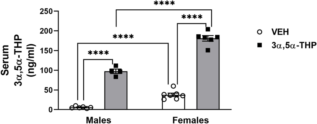
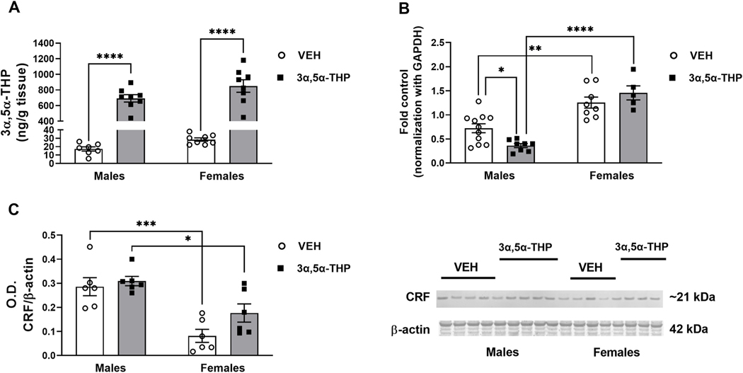
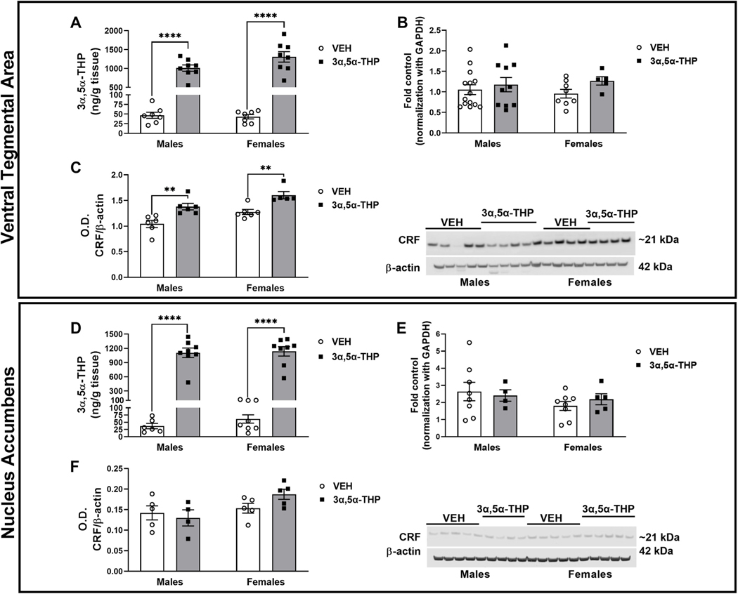
CRF is the main activator of the hypothalamic-pituitary-adrenal (HPA) axis in response to stress. CRF neurons are found mainly in the hypothalamus, but CRF positive cells and CRF1 receptors are also found in extrahypothalamic structures, including amygdala (CeA), hippocampus, NAc and VTA. CRF release in the hypothalamus is regulated by inhibitory GABAergic interneurons and extrahypothalamic glutamatergic inputs, and disruption of this balance is found in stress-related disorders and addiction. (3α,5α)3-hydroxypregnan-20-one (3α,5α-THP), the most potent positive modulator of GABAA receptors, attenuates the stress response reducing hypothalamic CRF mRNA expression and ACTH and corticosterone serum levels. In this study, we explored 3α,5α-THP regulation of hypothalamic and extrahypothalamic CRF mRNA and peptide expression, in male and female Sprague Dawley rats, following vehicle or 3α,5α-THP administration (15 mg/kg). In the hypothalamus, we found sex differences in CRF mRNA expression (females +74%, p < 0.01) and CRF peptide levels (females -71%, p < 0.001). 3α,5α-THP administration reduced hypothalamic CRF mRNA expression only in males (-50%, p < 0.05) and did not alter CRF peptide expression in either sex. In hippocampus and CeA, 3α,5α-THP administration reduced CRF peptide concentrations only in the male (hippocampus -29%, p < 0.05; CeA -62%, p < 0.01). In contrast, 3α,5α-THP injection increased CRF peptide concentration in the VTA of both males (+32%, p < 0.01) and females (+26%, p < 0.01). The results show sex and region-specific regulation of CRF signals and the response to 3α,5α-THP administration. This data may be key to successful development of therapeutic approaches for stress-related disorders and addiction.

Keywords: 3α,5α-THP; CRF; Extrahypothalamic CRF; HPA axis; Sex differences.


Conflict of interest statement

Declaration of competing interest

The authors have no conflicts of interest to declare.

Figures

Fig. 1.
Fig. 1.. Serum 3α,5α-THP levels are higher in female vs male rats following vehicle and 3α,5α-THP administration.
As expected, female rats showed higher levels of circulating 3α,5α-THP (males = 5.703 ± 1.02 ng/ml vs females = 37.59 ± 4.2 ng/ml, p = 0.0006). Following 3α,5α-THP (15 mg/kg) IP administration, plasma 3α,5α-THP levels were increased in both male and female rats (males VEH = 5.703 ± 1.02 ng/ml vs 3α,5α-THP = 97.46 ± 5.8 ng/ml, p < 0.0001; females VEH = 37.59 ± 4.2 ng/ml vs 3α,5α-THP = 181 ± 7 ng/ml, p < 0.0001); this increase was significantly different between male and female rats (males = 91.76 ± 5.8 ng/ml vs females = 143.41 ± 7 ng/ml; t (8) = 14.77, p < 0.001) (n = 4–7 per group). Significant effect was found using Two-way ANOVA, followed by Tukey HSD test, ****p < 0.0001. Data are represented as mean ± SEM. VEH = rats treated with vehicle; 3α,5α-THP = rats treated with 3α,5α-THP.
Fig. 2.
Fig. 2.. Sex differences in CRF signals after 3α,5α-THP administration in hypothalamus.
A. 3α,5α-THP (15 mg/kg) IP administration increased 3α,5α-THP levels in hypothalamus of male and female rats (males VEH = 17.29 ng/g tissue vs 3α,5α-THP = 692.2 ± 47 ng/g tissue, p < 0.0001; females VEH = 28.5 ± 2 ng/g tissue vs 3α,5α-THP = 850.8 ± 80.6 ng/g tissue, p < 0.0001) (n = 7–8 rats per group). B. The results of the qPCR analysis showed higher hypothalamic baseline CRF mRNA in female rats (males = 0.7224 ± 0.09 vs females = 1.255 ± 0.11, p = 0.0018. 3α,5α-THP injection decreased CRF mRNA only in male rats (males VEH = 0.7224 ± 0.09 vs 3α,5α-THP = 0.3624 ± 0.04, p = 0.0461) (n = 5–11 rats per group); after the treatment, females mRNA expression was higher than males (males = 0.3624 ± 0.04 vs females = 1.457 ± 0.15, p < 0.0001). C. In contrast to the mRNA results, in basal condition western blotting showed in female rats have lower hypothalamic CRF peptide levels than males (males = 0.2857 ± 0.04 vs females = 0.0816 ± 0.03, p = 0.0009); 3α,5α-THP injection did not affect the basal levels of CRF peptide, but male rats treated with 3α,5α-THP resulted in higher CRF levels than the same group of female rats (males = 0.3091 ± 0.02 vs females = 0.1766 ± 0.04, p = 0.0343) (n = 6 per group). Significant effect was found using Two-way ANOVA, followed by Tukey HSD test, *p < 0.05, **p < 0.01, ***p < 0.001, ****p < 0.0001. Data are represented as mean ± SEM. VEH = rats treated with vehicle; 3α,5α-THP = rats treated with 3α,5α-THP.
Fig. 3.
Fig. 3.. 3α,5α-THP administration decreased CRF peptide in hippocampus and central nucleus of amygdala of male rats.
Hippocampus: A. 3α,5α-THP (15 mg/kg) IP administration increased 3α,5α-THP levels in hippocampus of male and female rats (males VEH = 14.52 ± 1.8 ng/g tissue vs 3α,5α-THP = 938.3 ± 128.2 ng/g tissue, p < 0.0001; females VEH = 26.08 ± 4 ng/g tissue vs 3α,5α-THP = 1112 ± 138.3 ng/g tissue, p < 0.0001) (n = 6–8 rats per group). B. qPCR analysis did not show any sex or treatment differences in CRF mRNA expression (n = 6–14 rats per group). C. Western blotting analysis did not show any sex differences in baseline CRF peptide levels; however, 3α,5α-THP injection reduced CRF protein expression in male (VEH CRF ratio = 1.12 vs 3α,5α-THP CRF ratio = 0.7906, p = 0.0254), but not in female rats. Male rats treated with 3α,5α-THP exhibited lower CRF levels than the same group of female rats (male = 0.7906 ± 0.06 vs. female = 1.273 ± 0.05, p = 0.0017) (n = 5–6 per group). Central nucleus of amygdala: D. 3α,5α-THP (15 mg/kg) IP administration increased 3α,5α-THP levels in male and female rats, but the increase in female rats is higher than male rats (males VEH = 90.48 ± 37.24 ng/g tissue vs 3α,5α-THP = 778.1 ± 51.39 ng/g tissue, p < 0.0001; females VEH = 22.4 ± 1.96 ng/g tissue vs 3α,5α-THP = 1011 ± 95.73 ng/g tissue, p < 0.0001; males 3α,5α-THP = 778.1 ± 51.39 ng/g tissue vs females 3α,5α-THP = 1011 ± 95.73 ng/g tissue, p = 0.0386) (n = 6–8 rats per group). E. qPCR analysis did not show any sex or treatment differences in CRF mRNA expression (n = 6–14 rats per group). F. The results of western blotting analysis did not show any sex differences in CRF peptide levels under basal condition. However, 3α,5α-THP injection significantly reduced CRF protein expression in male (males VEH = 0.1658 ± 0.01 vs 3α,5α-THP = 0.0632 ± 0.02, p = 0.0033), but not in female rats. Male rats treated with 3α,5α-THP exhibited lower CRF levels than female rats (males = 0.0632 ± 0.02 vs females = 17.36 ± 0.02, p = 0.0017) (n = 5–6 per group). Significant effect was found using Two-Way ANOVA, followed by Tukey HSD test, *p < 0.05, **p < 0.01, ****p < 0.0001. Data are represented as mean ± SEM. VEH = rats treated with vehicle; 3α,5α-THP = rats treated with 3α,5α-THP.
Fig. 4.
Fig. 4.. 3α,5α-THP administration increased CRF peptide levels in ventral tegmental area (VTA), while did not affect CRF signaling in nucleus accumbens (NAc).
Ventral Tegmental Area: A. 3α,5α-THP (15 mg/kg) IP administration increased 3α,5α-THP levels in VTA of male and female rats (males VEH = 46.83 ± 7.94 ng/g tissue vs 3α,5α-THP = 1011 ± 83.39 ng/g tissue, p < 0.0001; females VEH = 43.11 ± 5.7 ng/g tissue vs 3α,5α-THP = 1308 ± 138.7 ng/g tissue, p < 0.0001) (n = 6–8 rats per group). B. qPCR analysis did not show any sex or treatment difference in CRF mRNA expression (n = 5–12 rats per group). C. Western blotting analysis showed an increase in both male and female rats following 3α,5α-THP injection (V males VEH = 1.043 ± 0.07 vs 3α,5α-THP = 1.380 ± 0.06, p = 0.0062; females VEH = 1.276 ± 0.05 vs females 3α,5α-THP = 1.602 ± 0.07, p = 0.0115) (n = 5–6 per group). Nucleus accumbens: D. 3α,5α-THP (15 mg/kg) IP administration increased 3α,5α-THP levels in male and female rats (males VEH = 36.78 ± 9.3 ng/g tissue vs 3α,5α-THP = 1105 ± 100.6 ng/g tissue, p < 0.0001; females VEH = 61.46 ± 14.51 ng/g tissue vs 3α,5α-THP = 1136 ± 100.7 ng/g tissue, p < 0.0001) (n = 6–8 rats per group). E. qPCR analysis did not show any sex or treatment differences in CRF mRNA expression (n = 4–8 rats per group). F. Western blotting analysis did not detect any change in CRF protein levels due by sex or treatment with 3α,5α-THP (n = 5–6 per group). Significant effect was found using Two-Way ANOVA, followed by Tukey HSD test, **p < 0.01, ****p < 0.0001. Data are represented as mean ± SEM. VEH = rats treated with vehicle; 3α,5α-THP = rats treated with 3α,5α-THP.

Similar articles

Cited by

References

    1. Aguirre A, Maturana CJ, Harcha PA, Saez JC, 2013. Possible involvement of TLRs and hemichannels in stress-induced CNS dysfunction via mastocytes, and glia activation. Mediat. Inflamm 2013, 893521 10.1155/2013/893521. - DOI - PMC - PubMed
    1. Akhter N, Madhoun A, Arefanian H, Wilson A, Kochumon S, Thomas R, Shenouda S, Al-Mulla F, Ahmad R, Sindhu S, 2019. Oxidative stress induces expression of the toll-like receptors (TLRs) 2 and 4 in the human peripheral blood mononuclear cells: implications for metabolic inflammation. Cell. Physiol. Biochem 53 (1), 1–18. 10.33594/000000117. - DOI - PubMed
    1. Balan I, Beattie MC, O’Buckley TK, Aurelian L, Morrow AL, 2019. Endogenous neurosteroid (3α,5α)3-hydroxypregnan-20-one inhibits toll-like-4 receptor activation and pro-inflammatory signaling in macrophages and brain. Sci. Rep 9 (1), 1220. 10.1038/s41598-018-37409-6. - DOI - PMC - PubMed
    1. Balan I, Warnock KT, Puche A, Gondre-Lewis MC, Aurelian L, 2018. Innately activated TLR4 signal in the nucleus accumbens is sustained by CRF amplification loop and regulates impulsivity. Brain Behav. Immun 69, 139–153. 10.1016/j.bbi.2017.11.008. - DOI - PMC - PubMed
    1. Bhattacharya A, Derecki NC, Lovenberg TW, Drevets WC, 2016. Role of neuro-immunological factors in the pathophysiology of mood disorders. Psychopharmacology (Berl) 233 (9), 1623–1636. 10.1007/s00213-016-4214-0. - DOI - PubMed

Publication types

Substances