. 2021 May:231:13-23.
doi: 10.1016/j.trsl.2021.01.005. Epub 2021 Jan 15.

Hemoglobin induces oxidative stress and mitochondrial dysfunction in oligodendrocyte progenitor cells

Affiliations

Hemoglobin induces oxidative stress and mitochondrial dysfunction in oligodendrocyte progenitor cells

Chirayu D Pandya et al. Transl Res. 2021 May.

Abstract

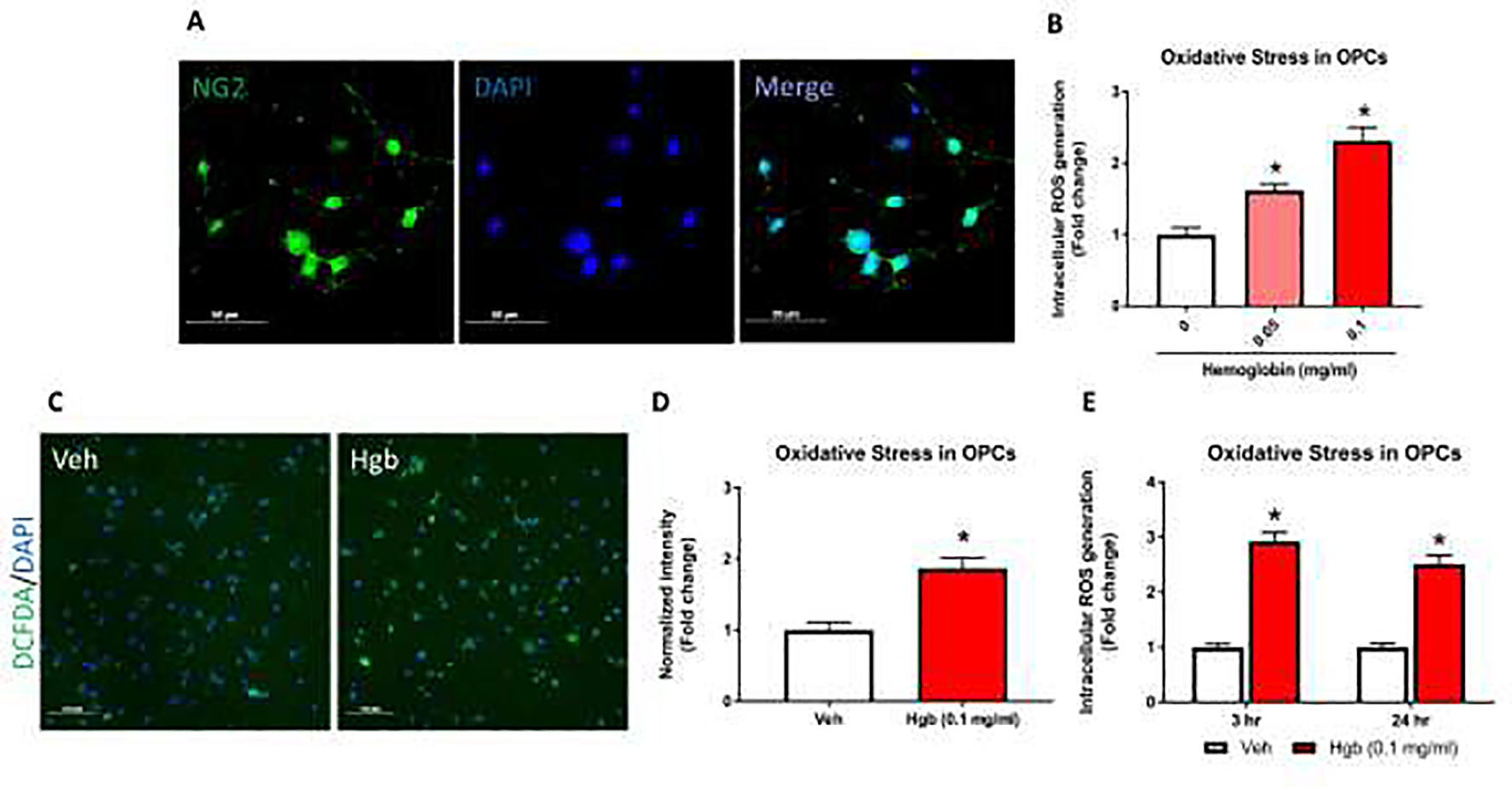
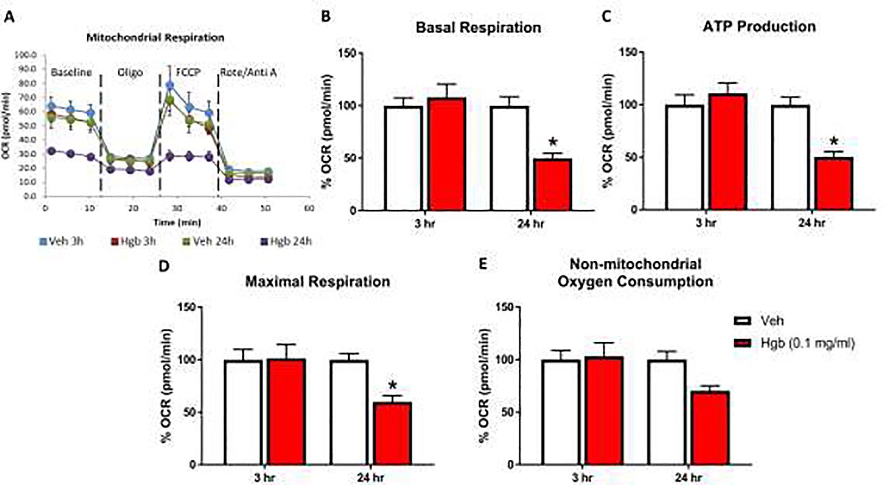
Oligodendrocyte progenitor cells (OPCs) in the infant brain give rise to mature oligodendrocytes that myelinate CNS axons. OPCs are particularly vulnerable to oxidative stress that occurs in many forms of brain injury. One common cause of infant brain injury is neonatal intraventricular hemorrhage (IVH), which releases blood into the CSF and brain parenchyma of preterm infants. Although blood contains the powerful oxidant hemoglobin, the direct effects of hemoglobin on OPCs have not been studied. We utilized a cell culture system to test if hemoglobin induced free radical production and mitochondrial dysfunction in OPCs. We also tested if phenelzine (PLZ), an FDA-approved antioxidant drug, could protect OPCs from hemoglobin-induced oxidative stress. OPCs were isolated from Sprague Dawley rat pups and exposed to hemoglobin with and without PLZ. Outcomes assessed included intracellular reactive oxygen species levels using 2',7'-dichlorodihydrofluorescein diacetate (DCF-DA) fluorescent dye, oxygen consumption using the XFe96 Seahorse assay, and proliferation measured by BrdU incorporation assay. Hemoglobin induced oxidative stress and impaired mitochondrial function in OPCs. PLZ treatment reduced hemoglobin-induced oxidative stress and improved OPC mitochondrial bioenergetics. The effects of hemoglobin and PLZ on OPC proliferation were not statistically significant, but showed trends towards hemoglobin reducing OPC proliferation and PLZ increasing OPC proliferation (P=0.06 for both effects). Collectively, our results indicate that hemoglobin induces mitochondrial dysfunction in OPCs and that antioxidant therapy reduces these effects. Therefore, antioxidant therapy may hold promise for white matter diseases in which hemoglobin plays a role, such as neonatal IVH.

Keywords: Free radicals; Intraventricular hemorrhage; Seahorse; White matter.


Conflict of interest statement

Declaration of Competing Interest The authors declare that there is no conflict of interest regarding the publication of this manuscript.

Figures

Fig 1.
Fig 1.. Hemoglobin-induced oxidative stress in primary OPCs.
Intracellular ROS measured by DCF-DA assay from isolated OPCs. (A) Immunofluorescence (IF) images show primary OPCs stained with NG2 (green) and the nuclear stain DAPI (blue). >90% of cells were NG2 positive. (B) OPCs were exposed to hemoglobin (0.05 and 0.1 mg/ml) for 24 hours and immediately assessed for intracellular ROS levels by DCF-DA via fluorescent microplate analysis. Both 0.05 and 0.1 mg/ml hemoglobin concentrations for 24 hours induced significant increase in ROS. (C) ROS production detected by DCF staining. Representative fluorescent images show that 24 hours of hemoglobin (Hgb) exposure increased ROS production in primary OPCs compared to vehicle (Veh). (D) Quantitative analyses show normalized DCF-DA fluorescent intensity. (E) Acute and chronic exposure of 0.1 mg/ml hemoglobin were tested for intracellular ROS generation. A significant increase oxidative stress was seen at 3 hours (2.92 fold) and 24 hours (2.51 fold) time point. Data are presented as mean ± SE; N = 3–8 for all groups. Each experiment was repeated two times and *=p < 0.05 was considered as significant vs vehicle.
Fig 2.
Fig 2.. PLZ protects the hemoglobin-induced changes in oxidative stress in OPCs.
Isolated OPCs were treated with Hgb for 3 hours and 24 hours with or without simultaneous PLZ treatment. ROS production were immediately determined by DCF-DA assay. PLZ treatment was able to protect OPCs ROS production in a time-dependent manner. (A) Different dose (10μM, 30 μM and 100 μM) of PLZ treatment at 10 μM for 3 hours found to be protective against hemoglobin exposed increased ROS levels. (B) PLZ treatment for 24 hours, at 10 μM shows protective trends against hemoglobin-induced ROS levels in OPCs. Data are presented as mean ± SE; N = 3 for all groups. Each experiment was repeated two times and *=p < 0.05 was considered as significant difference vs vehicle exposed group.
Fig 3.
Fig 3.. Hemoglobin decreases oxygen consumption rate (OCR) in OPCs.
Seahorse Cell Mito Stress Test was performed to measure OCR in acute (3 hours) and chronic (24 hours) incubation of vehicle and hemoglobin following a sequential addition of inhibitors of mitochondrial function such as oligomycin, carbonyl cyanide-ptrifluoromethoxyphenylhydrazone (FCCP), and a combination of rotenone and antimycin A; (A) OCR profile plot, (B) basal respiration, (C) ATP production, (D) maximal respiration and (E) non-mitochondrial consumption. Basal respiration was calculated after subtraction of non-mitochondrial respiration. ATP production or ATP-linked respiration were calculated following the addition of oligomycin. Maximal respiration was measured following the addition of FCCP. Data are presented as mean ± SE; N = 4 for all groups. Each experiment was repeated two times and *=p < 0.05 was considered as significant difference vs vehicle.
Fig 4.
Fig 4.. PLZ treatment enhance mitochondrial bioenergetics in hemoglobin exposed OPCs.
Oxygen consumption rate by seahorse were determined in primary OPCs after 24 hours of hemoglobin exposure in presence and absence of different concentration of PLZ; (A) OCR profile plot, (B) basal respiration, (C) ATP production, (D) maximal respiration and (E) non-mitochondrial consumption. PLZ at 10 μM concentration ameliorated the hemoglobin-induced decrease in basal and maximal respiration; however, ATP production shows increased trend. Non-mitochondrial respiration decreased with hemoglobin exposure, but did not changed with PLZ treatment. Data are presented as mean ± SE; N = 4–6 for all groups. Each experiment was repeated two times and *=p < 0.05 was considered significant vs vehicle.
Fig 5.
Fig 5.. PLZ enhances OPC proliferation.
Effects of hemoglobin (0.1 mg/ml) in presence and absence of PLZ (10 μM) treatment on OPC proliferation were measured by the BrdU incorporation assay. (A) Representative confocal images show that OPCs were stained with BrdU (red) and DAPI (blue) after 48 hours of PLZ and hemoglobin treatment. BrdU was added for the last 16 hours before fixing the cells. The images were acquired at 20X and scale bar is 100 μm. The pink spots represent the proliferative OPCs. (B) Quantitative analyses shows % proliferation, as calculated by BrdU+DAPI+ cells using HALO software. Data represents mean % proliferation ± SE (N = 3) and *P < 0.05 vs vehicle.
Fig 6.
Fig 6.
Schematic model of protective role of PLZ in hemoglobin-mediated ROS generation and altered mitochondrial bioenergetics in OPCs.

References

    1. Hart AR, Whitby EW, Griffiths PD, et al. Magnetic resonance imaging and developmental outcome following preterm birth: review of current evidence. Dev Med Child Neurol 2008;50:655–63. - PubMed
    1. Chen Z, Gao C, Hua Y, et al. Role of iron in brain injury after intraventricular hemorrhage. Stroke 2011;42:465–70. - PMC - PubMed
    1. Lee JY, Keep RF, He Y, et al. Hemoglobin and iron handling in brain after subarachnoid hemorrhage and the effect of deferoxamine on early brain injury. J Cereb Blood Flow Metab 2010;30:1793–803. - PMC - PubMed
    1. Goulding DS, Vogel RC, Pandya CD, et al. Neonatal hydrocephalus leads to white matter neuroinflammation and injury in the corpus callosum of Ccdc39 hydrocephalic mice. J Neurosurg Pediatr 2020:1–8. - PMC - PubMed
    1. Back SA. Perinatal white matter injury: the changing spectrum of pathology and emerging insights into pathogenetic mechanisms. Ment Retard Dev Disabil Res Rev 2006;12:129–40. - PubMed

Publication types