

Characterization of histological changes at the tillering stage (Z21) in resistant and susceptible wheat plants infected by Tilletia controversa Kühn
- PMID: 33461490
- PMCID: PMC7814547
- DOI: 10.1186/s12870-020-02819-0
Characterization of histological changes at the tillering stage (Z21) in resistant and susceptible wheat plants infected by Tilletia controversa Kühn
Abstract
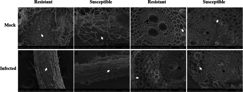
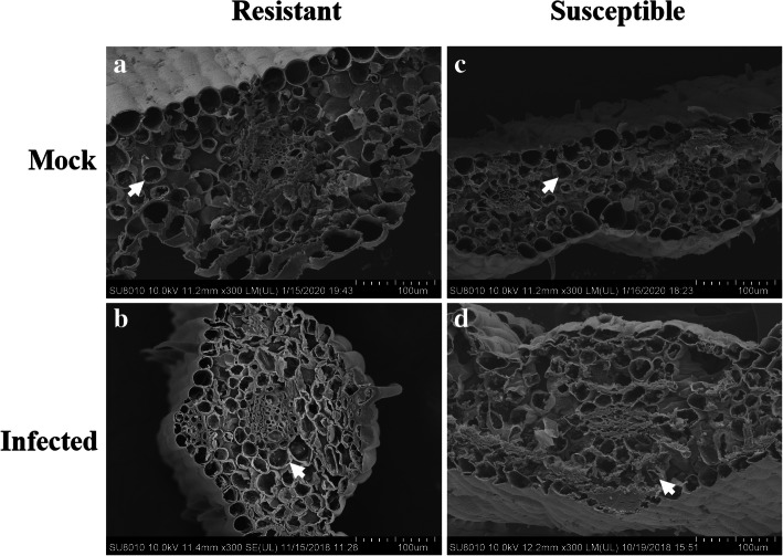
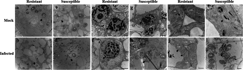
Background: Dwarf bunt, which is caused by Tilletia controversa Kühn, is a soilborne and seedborne disease that occurs worldwide and can lead to 70% or even total losses of wheat crops. However, very little information is available about the histological changes that occur in dwarf bunt-resistant and dwarf bunt-susceptible wheat plants at the tillering stage (Z21). In this study, we used scanning electron microscopy and transmission electron microscopy to characterize the histological changes at this stage in resistant and susceptible wheat cultivars infected by T. controversa.
Results: Using scanning electron microscopy, the root, stem, and leaf structures of resistant and susceptible cultivars were examined after T. controversa infection. The root epidermal and vascular bundles were more severely damaged in the susceptible T. controversa-infected plants than in the resistant plants. The stem cell and longitudinal sections were much more extensively affected in susceptible plants than in resistant plants after pathogen infection. However, slightly deformed mesophyll cells were observed in the leaves of susceptible plants. With transmission electron microscopy, we found that the cortical bundle cells and the cell contents and nuclei in the roots were more severely affected in the susceptible plants than in the resistant plants; in the stems and leaves, the nuclei, chloroplasts, and mesophyll cells changed significantly in the susceptible plants after fungal infection. Moreover, we found that infected susceptible and resistant plants were affected much more severely at the tillering stage (Z21) than at the seedling growth stage (Z13).
Conclusion: Histological changes in the wheat roots, stems and leaves were much more severe in T. controversa-infected susceptible plants than in infected resistant plants at the tillering stage (Z21).
Keywords: Histology; Scanning electron microscopy; Tilletia controversa Kühn; Transmission electron microscopy; Wheat dwarf bunt.
Conflict of interest statement
The authors declare that they have no competing interests.
Figures


Similar articles
-
Wheat Varietal Response to Tilletia controversa J. G. Kühn Using qRT-PCR and Laser Confocal Microscopy.Genes (Basel). 2021 Mar 16;12(3):425. doi: 10.3390/genes12030425. Genes (Basel). 2021. PMID: 33809560 Free PMC article.
-
Microbiome Signature of Endophytes in Wheat Seed Response to Wheat Dwarf Bunt Caused by Tilletia controversa Kühn.Microbiol Spectr. 2023 Feb 14;11(1):e0039022. doi: 10.1128/spectrum.00390-22. Epub 2023 Jan 10. Microbiol Spectr. 2023. PMID: 36625645 Free PMC article.
-
Characterization of the wheat cultivars against Tilletia controversa Kühn, causal agent of wheat dwarf bunt.Sci Rep. 2020 Jun 3;10(1):9029. doi: 10.1038/s41598-020-65748-w. Sci Rep. 2020. PMID: 32494028 Free PMC article.
-
Characterization of the microbial communities in wheat tissues and rhizosphere soil caused by dwarf bunt of wheat.Sci Rep. 2021 Mar 11;11(1):5773. doi: 10.1038/s41598-021-85281-8. Sci Rep. 2021. PMID: 33707584 Free PMC article.
-
Transcriptome analysis of wheat spikes in response to Tilletia controversa Kühn which cause wheat dwarf bunt.Sci Rep. 2020 Dec 9;10(1):21567. doi: 10.1038/s41598-020-78628-0. Sci Rep. 2020. PMID: 33299089 Free PMC article.
Cited by
-
Metabolomic Analysis of Wheat Grains after Tilletia laevis Kühn Infection by Using Ultrahigh-Performance Liquid Chromatography-Q-Exactive Mass Spectrometry.Metabolites. 2022 Aug 28;12(9):805. doi: 10.3390/metabo12090805. Metabolites. 2022. PMID: 36144210 Free PMC article.
-
Toward Understanding the Molecular Recognition of Fungal Chitin and Activation of the Plant Defense Mechanism in Horticultural Crops.Molecules. 2021 Oct 28;26(21):6513. doi: 10.3390/molecules26216513. Molecules. 2021. PMID: 34770922 Free PMC article. Review.
-
Characteristics of the Infection of Tilletia laevis Kühn (syn. Tilletia foetida (Wallr.) Liro.) in Compatible Wheat.Plant Pathol J. 2021 Oct;37(5):437-445. doi: 10.5423/PPJ.OA.05.2021.0082. Epub 2021 Oct 1. Plant Pathol J. 2021. PMID: 34847630 Free PMC article.
-
Understanding the Rice Fungal Pathogen Tilletia horrida from Multiple Perspectives.Rice (N Y). 2022 Dec 16;15(1):64. doi: 10.1186/s12284-022-00612-1. Rice (N Y). 2022. PMID: 36522490 Free PMC article. Review.
-
Kernel Transcriptome Profiles of Susceptible Wheat Genotypes in Response to Wheat Dwarf Bunt.Int J Mol Sci. 2023 Dec 8;24(24):17281. doi: 10.3390/ijms242417281. Int J Mol Sci. 2023. PMID: 38139108 Free PMC article.
References
-
- Young PA. A new variety of Tilletia tritici in Montana. Phytopathology. 1935;25:1921–1922.
-
- Yuan Q, Nian SJ, Yin YP, Li MH, Cai J, Wang ZK. Development of a PCR-based diagnostic tool specific to wheat dwarf bunt, caused by Tilletia controversa. Eur J Plant Pathol. 2009;124:585–594. doi: 10.1007/s10658-009-9445-z. - DOI
-
- Wang Y, Yu XX. Study on the incidence of Tilletia controversa Kühn in the absence of snow. Plant Quarantine. 1996;110:139–141.
Publication types
MeSH terms
Supplementary concepts
LinkOut - more resources
Full Text Sources
Other Literature Sources
