

Identification and molecular characterization of Subramaniula asteroides causing human fungal keratitis: a case report
- PMID: 33461505
- PMCID: PMC7814578
- DOI: 10.1186/s12879-021-05768-7
Identification and molecular characterization of Subramaniula asteroides causing human fungal keratitis: a case report
Abstract
Background: Keratitis due to by filamentous fungi are not easy to diagnose thus causing a delay in correct therapy. There are many descriptions of keratitis due to Candida, Fusarium and Aspergillus genera. Subramaniula genus has only recently been reported to cause human infections and there are few descriptions of eye infections due to this filamentous fungus. Diagnosis of fungal keratitis is usually based on microscopic and cultural techniques of samples obtained by corneal swabbing or scraping. Considering the amount of time required to obtain culture results it is wise to use other diagnostic methods, such as molecular analyses. Therapeutic options against these fungi are limited by low tissue penetration in the eye due to ocular barriers. We describe the first case of S. asteroides human keratitis treated with isavuconazole.
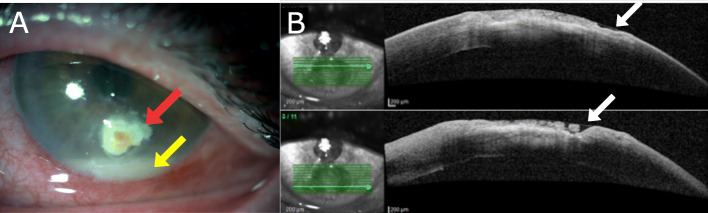
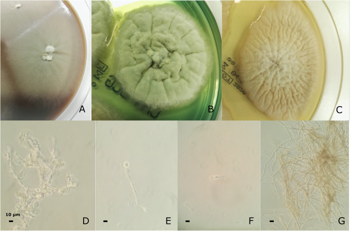
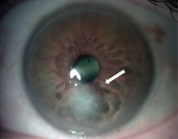
Case presentation: We describe a rare case of fungal keratitis unresponsive to antimicrobial treatment in a 65-year-old male patient without a history of diabetes or immunological diseases. He reported that the onset of symptoms occurred during a long holiday in Cape Verde Island. Initial treatment with topical antibiotics associated to steroids were ineffective, allowing a slow clinical progression of disease to corneal perforation. On admission in our Hospital, slit-lamp examination of the left eye showed conjunctival congestion and hyperemia, a large inferior corneal ulceration with brown pigment, corneal edema, about 3 mm of hypopyon and irido-lenticular synechiae. The slow clinical progression of the disease to corneal perforation and the aspect of the ulcer were consistent with a mycotic etiology. Molecular methods used on fungal colonies isolated by Sabouraud's dextrose agar cultures allowed the identification of Subramaniula asteroids from corneal scraping. Antimicrobial test showed a good susceptibility of this filamentous fungus to voriconazole and isavuconazole. Moreover, this fungal keratitis was successfully treated with isavuconazole, without side effects, observing a progressive clinical improvement.
Conclusions: Molecular methods may be useful for the identification of filamentous fungal keratitis on scraping samples thus shortening the time of diagnosis. Systemic therapy by isavuconazole could be useful to treat the filamentous fungal keratitis, reducing the possible adverse effects due to the use of voriconazole by systemic administration.
Keywords: Case report; Fungal keratitis; Isavuconazole; Molecular identification; Subramaniula asteroides; β-Tubulin gene.
Conflict of interest statement
The authors declare that they have no competing interests
Figures
Similar articles
-
Keratitis resulting from Thielavia subthermophila Mouchacca.Cornea. 2009 Oct;28(9):1067-9. doi: 10.1097/ICO.0b013e31819717f4. Cornea. 2009. PMID: 19724200
-
Recalcitrant Beauveria bassiana keratitis: confocal microscopy findings and treatment with posaconazole (Noxafil).Cornea. 2007 Sep;26(8):1008-10. doi: 10.1097/ICO.0b013e3180de4953. Cornea. 2007. PMID: 17721308
-
[Fungal keratitis caused by Scedosporium apiospermum: first report from Turkey].Mikrobiyol Bul. 2013 Oct;47(4):727-33. doi: 10.5578/mb.5262. Mikrobiyol Bul. 2013. PMID: 24237443 Turkish.
-
Cladosporium keratitis - a case report and literature review.BMC Ophthalmol. 2015 Aug 19;15:106. doi: 10.1186/s12886-015-0092-1. BMC Ophthalmol. 2015. PMID: 26286482 Free PMC article. Review.
-
Initial Case Report of Cladorrhinum samala Mycotic Keratitis.Cornea. 2022 Oct 1;41(10):1302-1304. doi: 10.1097/ICO.0000000000002992. Epub 2022 Jan 25. Cornea. 2022. PMID: 36107849 Review.
Cited by
-
New Biocomposite Electrospun Fiber/Alginate Hydrogel for Probiotic Bacteria Immobilization.Materials (Basel). 2021 Jul 10;14(14):3861. doi: 10.3390/ma14143861. Materials (Basel). 2021. PMID: 34300780 Free PMC article.
-
Morphological, Molecular and Metabolic Characterization of the Pigmented Fungus Subramaniula asteroids.J Fungi (Basel). 2022 Oct 29;8(11):1149. doi: 10.3390/jof8111149. J Fungi (Basel). 2022. PMID: 36354915 Free PMC article.
-
Pseudonectria keratitis-emerging pathogenic fungi in the eye.Ann Clin Microbiol Antimicrob. 2024 Jul 18;23(1):64. doi: 10.1186/s12941-024-00723-1. Ann Clin Microbiol Antimicrob. 2024. PMID: 39026348 Free PMC article.
-
Nanomedicine in fungal keratitis: current applications and future prospects.Front Microbiol. 2025 Jul 9;16:1618046. doi: 10.3389/fmicb.2025.1618046. eCollection 2025. Front Microbiol. 2025. PMID: 40703245 Free PMC article. Review.
-
An Atypical Etiology of Fungal Keratitis Caused by Roussoella neopustulans.J Fungi (Basel). 2022 May 15;8(5):507. doi: 10.3390/jof8050507. J Fungi (Basel). 2022. PMID: 35628762 Free PMC article.
References
Publication types
MeSH terms
Substances
Supplementary concepts
LinkOut - more resources
Full Text Sources
Other Literature Sources
