Review
. 2021 Jan 18;20(1):18.
doi: 10.1186/s12943-020-01263-w.

The role of m6A, m5C and Ψ RNA modifications in cancer: Novel therapeutic opportunities

Affiliations
Review

The role of m6A, m5C and Ψ RNA modifications in cancer: Novel therapeutic opportunities

Paz Nombela et al. Mol Cancer. .

Abstract

RNA modifications have recently emerged as critical posttranscriptional regulators of gene expression programmes. Significant advances have been made in understanding the functional role of RNA modifications in regulating coding and non-coding RNA processing and function, which in turn thoroughly shape distinct gene expression programmes. They affect diverse biological processes, and the correct deposition of many of these modifications is required for normal development. Alterations of their deposition are implicated in several diseases, including cancer. In this Review, we focus on the occurrence of N6-methyladenosine (m6A), 5-methylcytosine (m5C) and pseudouridine (Ψ) in coding and non-coding RNAs and describe their physiopathological role in cancer. We will highlight the latest insights into the mechanisms of how these posttranscriptional modifications influence tumour development, maintenance, and progression. Finally, we will summarize the latest advances on the development of small molecule inhibitors that target specific writers or erasers to rewind the epitranscriptome of a cancer cell and their therapeutic potential.

Keywords: 5-methylcytosine; Anti-cancer therapy; Cancer; Epitranscriptome; Inhibitors; Migration; N6-methyladenosine; Proliferation; Pseudouridine; RNA modifications; m5C; m6A; Ψ.


Conflict of interest statement

The authors declare that they have no competing interests.

Figures

Fig. 1
Fig. 1
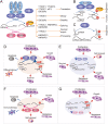
Schematic diagram of the location of m6A, m6Am, m5C and Ψ modifications on mRNA. Blue ribbon represents the mRNA with m7G-cap and a poly(A) tail. ATG and STOP codons are indicated. The writers, erasers, readers and function are listed in the text box attached to each modification. Modifications: m6A, 6-methyladenosine; m6Am, N6,2ʹ-O-dimethyladenosine; m5C, 5-methylcytosine; Ψ, pseudouridine. Proteins: METTL, methyltransferase-like; FTO, fat mass and obesity-associated protein; PUS, pseudouridine synthase; NSUN, NOL1/NOP2/SUN domain family member; ALyREF, Aly/REF Export Factor; ZCCHC4, zinc-finger CCHC domain-containing protein 4; ALKBH5, Alpha-Ketoglutarate-Dependent Dioxygenase AlkB Homolog 5; YTHDC, YTH domain-containing; YTHDF, YTH domain-containing family
Fig. 2
Fig. 2
Molecular mechanism of m6A deposition in RNA, biological function and implications in human cancer. a-c m6A RNA methylation landscape in mRNA (a) and ncRNA (b & c) mediated by writers (blue balloons), including METTL3, METTL14, WTAP, RBM15B, KIAA1429, ZC3H13 and METTL16, erasers (pink balloons) FTO and ALKBH5 and reader proteins (yellow balloons) YTHDF1, YTHDF2, YTHDF3, YTHDC1, YTHDC2 and HNRNPC, HNRNPG, HNRNPA2, HNRNPB1, IGF2BPs and eIF3, and their role in mRNA metabolism. d-g Aberrant m6A deposition in mRNA and ncRNAs promotes or suppresses tumour progression through METTL3/METTL14 upregulation (d) or downregulation (e) and FTO/ALKBH5 upregulation (f) or downregulation (g). Red arrows indicate induction. Blue arrows with flat end represent inhibition
Fig. 3
Fig. 3
Molecular mechanism of m5C deposition in RNA, biological function implication in human cancer. a-d m5C RNA deposition landscape in mRNA (a), tRNA (b), rRNA (d), and ncRNAs (c) mediated by writers (blue balloons) NSUN2–6, DNMT2 and NOP2, erasers (pink balloons) TET and ALKBH1, and reader proteins (yellow balloons) YBX1 and AlyREF, and their role in mRNA metabolism. e-h Aberrant m5C deposition in RNAs promotes or suppresses tumour progression through NSUN2 upregulation in mRNA, miRNA and lncRNA (e), NSUN2 downregulation in tRNA (f), NOP upregulation in rRNA (g) or NSUN5 downregulation in rRNA (h). Red arrows indicate induction. Blue arrows with flat end represent inhibition
Fig. 4
Fig. 4
Simplified structure of a tRNA and all known Ψ and m5C events in humans. Ψ modification is shaded in blue and m5C in purple. The table on the right indicates the position and modification in tRNA, the human enzyme that has been identified to catalyse this modification, the sub-cellular localization of tRNAs containing the modification. m5C, 5-methylcytosine; f5C, 5-formylcytosine Ψ, pseudouridine; Ψ derivates: Ψm, 2ʹ-O-methylpseudouridine and m1Ψ, 1-methylpseudouridine. Proteins: PUS, pseudouridine synthase; RPUSD, RNA pseudouridine synthase domain-containing protein; NSUN, NOL1/NOP2/SUN domain family member; DNMT2, DNA methyltransferase 2. f5C* is formed from m5C by ALKBH1 (Alpha-Ketoglutarate-Dependent Dioxygenase AlkB Homolog 1) in mitochondrial tRNAs. Ψ derivates are formed from Ψ by methyltransferases
Fig. 5
Fig. 5
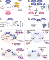
Ψ deposition mechanisms and pathological implications in cancer. a Schematic illustration of RNA-dependent pseudouridylation mechanisms. Substrate recognition is achieved by sequence and structure homology of the substrate with the structural stems and loops formed by box H/ACA small nucleolar RNAs (snoRNA). Dyskerin (DKC1) is the catalytic unit and non-histone protein 2 (Nhp2), nucleolar protein 10 (Nop10) and glycine-arginine-rich protein 1 (Gar1) are regulatory units. The RNA substrate is represented in green. b Illustration of RNA-independent pseudouridylation mechanisms. Substrate recognition and catalysis are performed by one single pseudouridine synthases (PUS) (blue). c Representative scheme illustrating the target specificity for each pseudouridine synthases. The fate of each modified RNA is also illustrated with orange arrows. Pseudouridylated RNAs may be also recognised by still unknown readers (?). d Altered expression of pseudouridine synthases can lead to cancer. For example, reduced expression of DKC1 induces a reduction of pseudouridylation in TERC and rRNA, leading to dysfunctional TERC and rRNA and increasing tumourigenesis. e In glioma, An increased expression of DKC1 can lead to an increased Ψ deposition on rRNA, snRNA and TERC, and thus promoting cancer cell growth and migration. f PUS7 decreased expression leads to hypomodified tRNA-derived fragments, leading to increased self-renewal and survival in bone marrow mononuclear cells, promoting tumourigenesis. Red arrows indicate an increase and blue arrows a decrease of processes or enzymes

References

    1. Boccaletto P, Machnicka MA, Purta E, Piatkowski P, Baginski B, Wirecki TK, de Crecy-Lagard V, Ross R, Limbach PA, Kotter A, et al. MODOMICS: a database of RNA modification pathways. 2017 update. Nucleic Acids Res. 2018;46:D303–D307. doi: 10.1093/nar/gkx1030. - DOI - PMC - PubMed
    1. Cohn WE. Pseudouridine, a carbon-carbon linked ribonucleoside in ribonucleic acids: isolation, structure, and chemical characteristics. J Biol Chem. 1960;235:1488–1498. doi: 10.1016/S0021-9258(18)69432-3. - DOI - PubMed
    1. Barbieri I, Kouzarides T. Role of RNA modifications in cancer. Nat Rev Cancer. 2020;20:303–322. doi: 10.1038/s41568-020-0253-2. - DOI - PubMed
    1. Jonkhout N, Tran J, Smith MA, Schonrock N, Mattick JS, Novoa EM. The RNA modification landscape in human disease. RNA. 2017;23:1754–1769. doi: 10.1261/rna.063503.117. - DOI - PMC - PubMed
    1. Davalos V, Blanco S, Esteller M. SnapShot: Messenger RNA Modifications. Cell. 2018;174:498–498 e491. doi: 10.1016/j.cell.2018.06.046. - DOI - PubMed

Publication types