. 2021 Jan 18;19(1):23.
doi: 10.1186/s12951-020-00762-8.

STED lithography in microfluidics for 3D thrombocyte aggregation testing

Affiliations

STED lithography in microfluidics for 3D thrombocyte aggregation testing

Bianca Buchegger et al. J Nanobiotechnology. .

Abstract

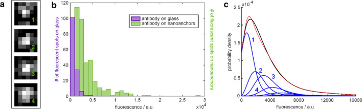
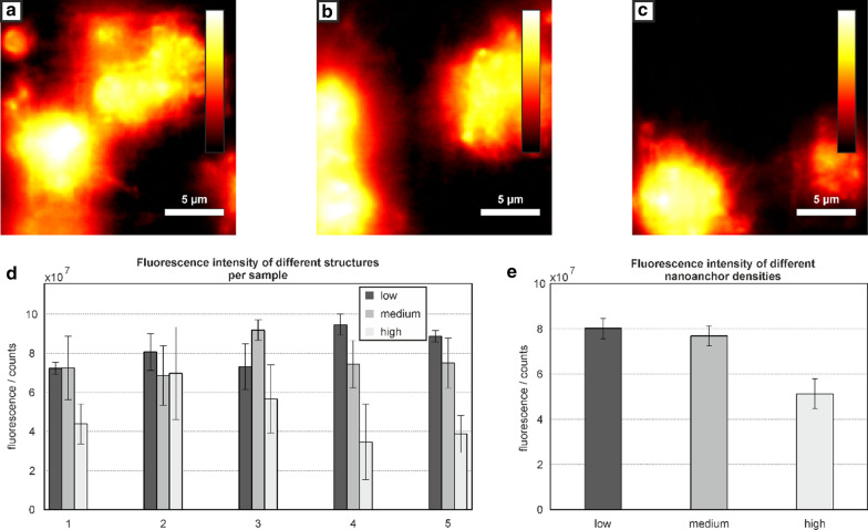
Three-dimensional photopolymerization techniques such as multiphoton polymerization lithography (MPL) and stimulated emission depletion (STED) lithography are powerful tools for fabricating structures in the sub-µm range. Combining these techniques with microfluidics enables us to broaden the range of their applications. In this study, we show a microfluidic device enhanced with MPL structures carrying STED-lithographically written nanoanchors that promote binding of the von Willebrand factor (vWF). The density of vWF is adjusted by varying the number of the nanoanchors on the 3D structures. This allows us to study the impact of the density of vWF on the activation of thrombocytes. The activation of the thrombocytes seems to decrease with the density of vWF on the 3D scaffolds inside the microfluidic channels.

Keywords: Microfluidics; Multiphoton polymerization lithography; Stimulated emission depletion lithography; Thrombocyte activation; Von Willebrand factor.


Conflict of interest statement

The authors declare that they have no competing interests.

Figures

Fig. 1
Fig. 1
Microfluidic device for thrombocyte activation studies. a Sketch representing side- and top-view of the microfluidic device. Blue: glass slides, orange: double-sided adhesive tape with a thickness of 83.1 µm, green: in- and outlet connectors. b Scanning electron microscopy (SEM) images of the structures inside the microfluidic channel. Protein repellent MPL grids on posts (grey) carry STED-lithography written nanoanchors (green). c Left: Sketch of the protein repellent structure (grey) with protein binding nanoanchors (green). Right: Fluorescence microscopy image of an empty scaffold. Excitation wavelength: 491 nm, illumination time 5 ms. d Left: Sketch of the experiment for thrombocyte activation. The protein binding nanoanchors were labeled with von Willebrand factor (vWF) molecules. Subsequently, thrombocytes were added to the microfluidic device. Fluorescently labeled antibodies (a-CD62P Alexa 647) were bound to the activated thrombocytes. Right: Fluorescence image of activated thrombocytes bound on top of the scaffolds. Excitation wavelength: 642 nm, illumination time 5 ms. e Left: Sketch of the control experiment where vWF molecules were omitted. The structures were incubated with a thrombocyte concentrate under flow conditions and subsequently incubated with fluorescently labeled a-CD62p antibodies. Right: There were no activated thrombocytes present on the structures. Excitation wavelength: 642 nm, illumination time 5 ms
Fig. 2
Fig. 2
Estimation of the number of vWF molecules on nanoanchors. a Examples of fluorescence peaks on nanoanchors carrying most probably (from top to bottom) one (809 counts), two (1527 counts), three (2563 counts), and four (3306 counts) fluorescent IgG antibodies. b Distribution of Alexa647 fluorescence signals (a.u.) from anti-vWF IgGs. Purple: single antibody signals originating from sparsely and randomly distributed antibodies on a piranha-cleaned glass surface; green: fluorescence signals from nanoanchors loaded with vWF. Illumination time: 5 ms. c Black line: Probability density function fitted to the experimental signals from anti-vWF IgGs on the nanoanchors. The blue curve 1 is a weighted fit of the measured probability density function for sparsely distributed IgGs on glass and blue curves 2, 3, 4 etc. are the weighted extrapolations for 2, 3, 4 etc. IgGs, respectively. The red curve is the sum of the blue curves. The weighting indicates that 34% of the nanoanchors carry exactly one antibody, 20% carry two, 21% carry three, 10% carry four and 15% carry five or more fluorescent IgG antibodies
Fig. 3
Fig. 3
Thrombocyte activation studies. ac Fluorescence images of activated thrombocytes on top of the 3D structures inside the microfluidic channels. The grids in ac carry a low, medium and high density of nanoanchors with integrated intensities of 5.17×10⁷ counts, 4.34×10⁷ counts and 2.54×10⁷ counts, respectively. d Fluorescence signal of anti-CD62p-Alexa®647, integrated in the proximity to each structure (picture section of 26.4 × 26.4 µm) for grids 1 to 5 in downstream direction. Each data point is averaged over four experiments. e Average fluorescence from the first four grids for the three experiments with low, medium and high density of nanoanchors

References

    1. Maruo S, Nakamura O, Kawata S. Three-dimensional microfabrication with two-photon-absorbed photopolymerization. Opt Lett. 1997;22:132–4. doi: 10.1364/OL.22.000132. - DOI - PubMed
    1. Kumi G, Yanez CO, Belfield KD, Fourkas JT. High-speed multiphoton absorption polymerization: fabrication of microfluidic channels with arbitrary cross-sections and high aspect ratios. Lab Chip. 2010;10:1057–60. doi: 10.1039/B923377F. - DOI - PubMed
    1. Lemma ED, Spagnolo B, de Vittorio M, Pisanello F. Studying Cell Mechanobiology in 3D: The Two-Photon Lithography Approach. Trends Biotechnol. 2019;37:358–72. doi: 10.1016/j.tibtech.2018.09.008. - DOI - PubMed
    1. Gansel JK, Thiel M, Rill MS, Decker M, Bade K, Saile V, et al. Gold helix photonic metamaterial as broadband circular polarizer. Science. 2009;325:1513–5. doi: 10.1126/science.1177031. - DOI - PubMed
    1. Gattass RR, Mazur E. Femtosecond laser micromachining in transparent materials. Nature Photon. 2008;2:219–25. doi: 10.1038/nphoton.2008.47. - DOI

LinkOut - more resources