

Topographic patterns of white matter hyperintensities are associated with multimodal neuroimaging biomarkers of Alzheimer's disease
- PMID: 33461618
- PMCID: PMC7814451
- DOI: 10.1186/s13195-020-00759-3
Topographic patterns of white matter hyperintensities are associated with multimodal neuroimaging biomarkers of Alzheimer's disease
Abstract
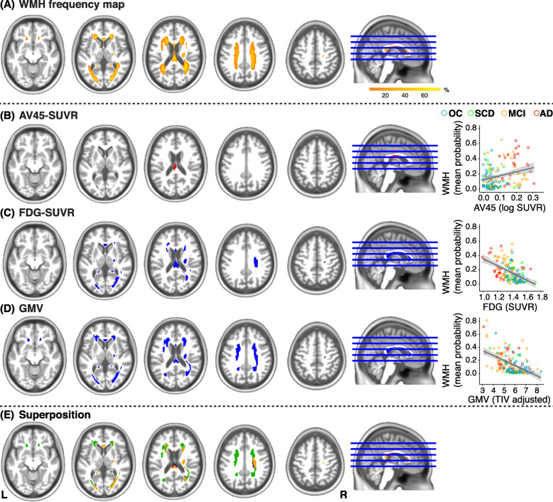
Background: White matter hyperintensities (WMH) are frequently found in Alzheimer's disease (AD). Commonly considered as a marker of cerebrovascular disease, regional WMH may be related to pathological hallmarks of AD, including beta-amyloid (Aβ) plaques and neurodegeneration. The aim of this study was to examine the regional distribution of WMH associated with Aβ burden, glucose hypometabolism, and gray matter volume reduction.
Methods: In a total of 155 participants (IMAP+ cohort) across the cognitive continuum from normal cognition to AD dementia, FLAIR MRI, AV45-PET, FDG-PET, and T1 MRI were acquired. WMH were automatically segmented from FLAIR images. Mean levels of neocortical Aβ deposition (AV45-PET), temporo-parietal glucose metabolism (FDG-PET), and medial-temporal gray matter volume (GMV) were extracted from processed images using established AD meta-signature templates. Associations between AD brain biomarkers and WMH, as assessed in region-of-interest and voxel-wise, were examined, adjusting for age, sex, education, and systolic blood pressure.
Results: There were no significant associations between global Aβ burden and region-specific WMH. Voxel-wise WMH in the splenium of the corpus callosum correlated with greater Aβ deposition at a more liberal threshold. Region- and voxel-based WMH in the posterior corpus callosum, along with parietal, occipital, and frontal areas, were associated with lower temporo-parietal glucose metabolism. Similarly, lower medial-temporal GMV correlated with WMH in the posterior corpus callosum in addition to parietal, occipital, and fontal areas.
Conclusions: This study demonstrates that local white matter damage is correlated with multimodal brain biomarkers of AD. Our results highlight modality-specific topographic patterns of WMH, which converged in the posterior white matter. Overall, these cross-sectional findings corroborate associations of regional WMH with AD-typical Aß deposition and neurodegeneration.
Keywords: Alzheimer’s disease; Alzheimer’s disease pathology; Cerebrovascular disease; White matter lesion.
Conflict of interest statement
Malo Gaubert reports no competing interests.
Catharina Lange reports no competing interests.
Antoine Garnier-Crussard reports no competing interests.
Theresa Köbe reports no competing interests.
Salma Bougacha reports no competing interests.
Julie Gonneaud reports no competing interests.
Robin de Flores reports no competing interests.
Clémence Tomadesso reports no competing interests.
Florence Mézenge reports no competing interests.
Brigitte Landeau reports no competing interests.
Vincent de la Sayette reports no competing interests.
Gaël Chételat reports no competing interests.
Miranka Wirth reports no competing interests.
Figures
References
-
- Wardlaw JM, Smith EE, Biessels GJ, Cordonnier C, Fazekas F, Frayne R, Lindley RI, O'Brien JT, Barkhof F, Benavente OR, et al. Neuroimaging standards for research into small vessel disease and its contribution to ageing and neurodegeneration. Lancet Neurol. 2013;12:822–838. doi: 10.1016/S1474-4422(13)70124-8. - DOI - PMC - PubMed
-
- Garnier-Crussard A, Bougacha S, Wirth M, Andre C, Delarue M, Landeau B, Mezenge F, Kuhn E, Gonneaud J, Chocat A, et al. White matter hyperintensities across the adult lifespan: relation to age, Abeta load, and cognition. Alzheimers Res Ther. 2020;12:127. doi: 10.1186/s13195-020-00669-4. - DOI - PMC - PubMed
Publication types
MeSH terms
Substances
LinkOut - more resources
Full Text Sources
Other Literature Sources
Medical
Miscellaneous
