. 2021 Mar:131:171-179.
doi: 10.1016/j.molimm.2020.12.035. Epub 2021 Jan 15.

TREM2 alters the phagocytic, apoptotic and inflammatory response to Aβ42 in HMC3 cells

Affiliations

TREM2 alters the phagocytic, apoptotic and inflammatory response to Aβ42 in HMC3 cells

Rumana Akhter et al. Mol Immunol. 2021 Mar.

Abstract

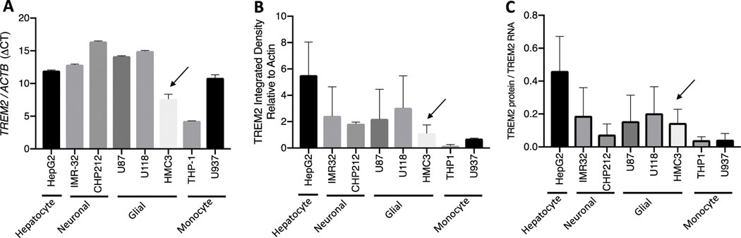
Alzheimer's disease (AD) is characterized by the accumulation in the brain of extracellular amyloid β (Aβ) plaques as well as intraneuronal inclusions (neurofibrillary tangles) consisting of total tau and phosphorylated tau. Also present are dystrophic neurites, loss of synapses, neuronal death, and gliosis. AD genetic studies have highlighted the importance of inflammation in this disease by identifying several risk associated immune response genes, including TREM2. TREM2 has been strongly implicated in basic microglia function including, phagocytosis, apoptosis, and the inflammatory response to Aβ in mouse brain and primary cells. These studies show that microglia are key players in the response to Aβ and in the accumulation of AD pathology. However, details are still missing about which apoptotic or inflammatory factors rely on TREM2 in their response to Aβ, especially in human cell lines. Given these previous findings our hypothesis is that TREM2 influences the response to Aβ toxicity by enhancing phagocytosis and inhibiting both the BCL-2 family of apoptotic proteins and pro-inflammatory cytokines. Aβ42 treatment of the human microglial cell line, HMC3 cells, was performed and TREM2 was overexpressed or silenced and the phagocytosis, apoptosis and inflammatory response were evaluated. Results indicate that a robust phagocytic response to Aβ after 24 h requires TREM2 in HMC3 cells. Also, TREM2 inhibits Aβ induced apoptosis by activating the Mcl-1/Bim complex. TREM2 is involved in activation of IP-10, MIP-1a, and IL-8, while it inhibits FGF-2, VEGF and GRO. Taken together, TREM2 plays a role in enhancing the microglial functional response to Aβ toxicity in HMC3 cells. This novel information suggests that therapeutic strategies that seek to activate TREM2 may not only enhance phagocytosis and inhibit apoptosis, but may also inhibit beneficial inflammatory factors, emphasizing the need to define TREM2-related inflammatory activity in not only mouse models of AD, but also in human AD.

Keywords: Apoptosis; Aβ; Inflammatory factors; Microglia; Phagocytosis; TREM2.


Conflict of interest statement

Declaration of Competing Interest

The authors declare no conflict of interest.

Figures

Fig 1.
Fig 1.. HMC3 cells express low to moderate TREM2 levels relative to other cell lines.
TREM2 RNA ΔCT levels (Relative to ACTB) are lower than most other cell lines (A). TREM2 protein (Relative to Actin B) are lower than most other cell lines (B). TREM2 protein relative to TREM2 RNA is lower the HepG2 cell line (C).
Fig 2.
Fig 2.. Aβ42 treatment of HMC3 cells increases TREM2.
Treatment of HMC3 cells with 5 μM Aβ for 24 hours increases cell surface TREM2 expression (A). TREM2 full length protein expression is significantly increase after 24 hour 5 μM Aβ42 treatment (25 kDa: p-value = 0.0247) (B). In a separate experiment, full length TREM2 relative to Actin (Western Blot: 25 kDa) is significantly increased, upon 24 hour 5 μM Aβ42 treatment while overexpression of TREM2 (OE) plus Aβ42 treatment does not significantly increase TREM2. Knockdown of TREM2 (siRNA) significantly depletes TREM2 with and without 24 hour 5 μM Aβ42 treatment (C). Fisher’s LSD test without correction for multiple comparisons significant p-value <0.05.
Fig 3.
Fig 3.. Lack of TREM2 decreases Aβ42 stimulated phagocytosis in HMC3 cells.
Treatment of HMC3 cells with 5 μM Aβ42 for 24 hours changes cell morphology from resting ramified microglial (A) to activated amoeboid microglia (B). Maximum phagocytosis is observed after 24 hours of 5 μM Aβ42 treatment (lines represent significant p- values <0.01) (C). After 24 hours of 5 μM Aβ42 treatment there is a significant increase in phagocytosis with or without overexpression of TREM2 and upon TREM2 knockdown there is a decrease in phagocytosis with or without 24 hour 5 μM Aβ42 treatment (D).
Fig 4.
Fig 4.. Aβ42 treatment alters cell survival, pro-apoptotic and anti-apoptotic factors in HMC3 cells.
HMC3 cell viability significantly declines after 8 hours of 5 μM Aβ42 treatment Bars represent Fisher’s LSD test without correction for multiple comparisons significant p-value <0.05.
Fig 5.
Fig 5.. TREM2 modulates apoptotic machinery in HMC3 cells.
(1) Bax, Bad, and pBad are reduced in response to 24 hour Aβ42 treatment. (2) TREM2 overexpression (TREM2 OE) does not influence these apoptotic proteins. (3) TREM2 silencing (TREM2 siRNA) increases Bax. (4) Mcl-1/Bim is increased in response to 24 hour Aβ42 treatment in the context of TREM2 overexpression. (5) Bax, Bcl-xL/Bad are decreased in response to 24 hour Aβ42 treatment in the context of TREM2 silencing. (6) In response to 24 hour Aβ42 treatment TREM2 silencing results in a decrease in Bax compared to TREM2 overexpression. (A) Significance (p-values) are Tukey adjusted for multiple comparisons and mean difference (Mean Diff.) is shown for each apoptotic factor. (B)
Fig 6.
Fig 6.. Proinflammatory factors generally increased upon TREM2 overexpression in HMC3 cells.
(1) No change in inflammatory factors after 24 hour Aβ42 treatment. (2) No change in inflammatory factors with TREM2 overexpression. (3) No change in inflammatory factors when TREM2 silenced. (4) No change in inflammatory factors with TREM2 overexpression after 24 hour Aβ42 treatment. (5) FGF-2 is increased and MIP- 1a is decreased when TREM2 silenced after 24 hour Aβ42 treatment. (6) In response to 24 hour Aβ42 treatment TREM2 silencing results in a decrease in IP-10, MIP-1a, IL- 8 and a increase in FGF-2, VEGF, GRO, compared to TREM2 overexpression. (A) Significance (p-values) are Tukey adjusted for multiple comparisons and mean difference (Mean Diff.) is shown for each apoptotic factor. (B)

References

    1. Abduljaleel Z, Al-Allaf FA, Khan W, Athar M, Shahzad N, Taher MM, Elrobh M, Alanazi MS and El-Huneidi W. (2014). “Evidence of trem2 variant associated with triple risk of Alzheimer’s disease.” PLoS One 9(3): e92648. - PMC - PubMed
    1. Afridi R, Lee WH and Suk K. (2020). “Microglia Gone Awry: Linking Immunometabolism to Neurodegeneration.” Front Cell Neurosci 14: 246. - PMC - PubMed
    1. Akhter R, Saleem S, Saha A and Biswas SC (2018). “The pro-apoptotic protein Bmf co-operates with Bim and Puma in neuron death induced by beta-amyloid or NGF deprivation.” Mol Cell Neurosci 88: 249–257. - PubMed
    1. Akhter R, Sanphui P and Biswas SC (2014). “The essential role of p53-up-regulated modulator of apoptosis (Puma) and its regulation by FoxO3a transcription factor in beta-amyloid-induced neuron death.” J Biol Chem 289(15): 10812–10822. - PMC - PubMed
    1. Akhter R, Sanphui P, Das H, Saha P and Biswas SC (2015). “The regulation of p53 up-regulated modulator of apoptosis by JNK/c-Jun pathway in beta-amyloid-induced neuron death.” J Neurochem 134(6): 1091–1103. - PubMed

Publication types

LinkOut - more resources