Initial presentation of lymphangioleiomyomatosis in third trimester of pregnancy
- PMID: 33462017
- PMCID: PMC7813302
- DOI: 10.1136/bcr-2020-237824
Initial presentation of lymphangioleiomyomatosis in third trimester of pregnancy
Abstract
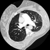
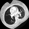
Lymphangioleiomyomatosis (LAM) is a progressive cystic lung disease which mostly affects premenopausal women and could be exacerbated by pregnancy. Therefore, it is thought that oestrogen plays an important role in LAM pathogenesis. Here, a case of LAM is described in which the first presentation of symptoms occurred during the third trimester of pregnancy. Symptoms included acute onset dyspnoea and chest pain at gestational age of 39 weeks and 2 days. A CT was performed which showed multiple thin-walled cysts and a small pneumothorax. Serum levels of vascular endothelial growth factor-D (VEGF-D) was 1200 pg/mL. The typical cystic lung changes on chest CT in combination with elevated VEGF-D is diagnostic for LAM. Given the risk of respiratory complications, the decision was made to deliver the baby at a gestational age of 39 weeks and 6 days by a planned caesarean section. Both mother and child were discharged home in good condition.
Keywords: gynaecology and fertility; obstetrics; pneumothorax; pregnancy; respiratory system.
© BMJ Publishing Group Limited 2020. No commercial re-use. See rights and permissions. Published by BMJ.
Conflict of interest statement
Competing interests: None declared.
Figures
Similar articles
-
Serum vascular endothelial growth factor-D as a diagnostic and therapeutic biomarker for lymphangioleiomyomatosis.PLoS One. 2019 Feb 28;14(2):e0212776. doi: 10.1371/journal.pone.0212776. eCollection 2019. PLoS One. 2019. PMID: 30818375 Free PMC article.
-
Diagnostic and Treatment Monitoring Potential of Serum Vascular Endothelial Growth Factor-D in Lymphangioleiomyomatosis.Lymphology. 2016 Sep;49(3):140-9. Lymphology. 2016. PMID: 29906075
-
Pulmonary lymphangioleiomyomatosis. A study of 69 patients. Groupe d'Etudes et de Recherche sur les Maladies "Orphelines" Pulmonaires (GERM"O"P).Medicine (Baltimore). 1999 Sep;78(5):321-37. doi: 10.1097/00005792-199909000-00004. Medicine (Baltimore). 1999. PMID: 10499073
-
Lymphangioleiomyomatosis and pregnancy: a mini-review.Arch Gynecol Obstet. 2024 Jun;309(6):2339-2346. doi: 10.1007/s00404-024-07478-2. Epub 2024 Apr 9. Arch Gynecol Obstet. 2024. PMID: 38594407 Free PMC article. Review.
-
Lymphangioleiomyomatosis: A review.Eur J Intern Med. 2008 Jul;19(5):319-24. doi: 10.1016/j.ejim.2007.10.015. Epub 2007 Dec 26. Eur J Intern Med. 2008. PMID: 18549932 Review.
Cited by
-
Lymphangioleiomyomatosis and Pregnancy-Do We Have All the Answers for a Woman Who Desires to Conceive?-Literature Review.Cancers (Basel). 2025 Jan 20;17(2):323. doi: 10.3390/cancers17020323. Cancers (Basel). 2025. PMID: 39858105 Free PMC article. Review.
-
Pathologically confirmed diffuse alveolar haemorrhage in lymphangioleiomyomatosis.BMJ Case Rep. 2021 Nov 9;14(11):e238713. doi: 10.1136/bcr-2020-238713. BMJ Case Rep. 2021. PMID: 34753716 Free PMC article.
-
Successful live birth via in vitro fertilization in a patient with lymphangioleiomyomatosis: a case report.J Med Case Rep. 2025 Aug 22;19(1):426. doi: 10.1186/s13256-025-05465-y. J Med Case Rep. 2025. PMID: 40847317 Free PMC article.
-
Complications of lymphangioleiomyomatosis in pregnancy: a case report and review of the literature.AJOG Glob Rep. 2024 Jan 10;4(1):100309. doi: 10.1016/j.xagr.2024.100309. eCollection 2024 Feb. AJOG Glob Rep. 2024. PMID: 38327672 Free PMC article.
-
The Epidemiology and Clinical Features of Lymphangioleiomyomatosis (LAM): A Descriptive Study of 33 Case Reports.Cureus. 2023 Aug 15;15(8):e43513. doi: 10.7759/cureus.43513. eCollection 2023 Aug. Cureus. 2023. PMID: 37719610 Free PMC article. Review.
References
-
- Factsheets LAM. Available: https://www.europeanlung.org/en/
Publication types
MeSH terms
LinkOut - more resources
Full Text Sources
Other Literature Sources
Medical


