A wide spectrum of rare clinical variants of Zinner syndrome
- PMID: 33462046
- PMCID: PMC7813347
- DOI: 10.1136/bcr-2020-239254
A wide spectrum of rare clinical variants of Zinner syndrome
Abstract
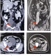
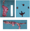
Congenital malformations of the seminal vesicles (SVs) are rare and are associated with abnormalities of the ipsilateral urinary tracts as embryologically both the ureteral buds and SVs arise from the mesonephric ducts. The triad of SV cysts, ipsilateral renal agenesis and ejaculatory duct obstruction is known as the Zinner syndrome. We, herein, present three very rare presentations of Zinner syndrome. Case 1 presented with haematuria, and was found to have a large SV cyst with stones and underwent a robotic cyst excision. Case 2 presented with primary infertility, and was found to have a variant of Zinner syndrome. Case 3 was a known case of chronic kidney disease on maintenance haemodialysis who presented with fever and oliguria. He was found to have Zinner syndrome and underwent aspiration of SV abscess. To the best of our knowledge, such varying presentations of Zinner syndrome have been rarely reported thus far.
Keywords: congenital disorders; haematuria; urological surgery; urology.
© BMJ Publishing Group Limited 2020. No commercial re-use. See rights and permissions. Published by BMJ.
Conflict of interest statement
Competing interests: None declared.
Figures


Similar articles
-
Multicystic seminal vesicle with ipsilateral renal agenesis: two cases of Zinner syndrome.Scand J Urol. 2017 Feb;51(1):81-84. doi: 10.1080/21681805.2016.1257650. Epub 2016 Dec 1. Scand J Urol. 2017. PMID: 27905212
-
A rare variant of zinner syndrome with ejaculatory duct cyst: case report and challenges in diagnosis and management.BMC Urol. 2024 Dec 4;24(1):263. doi: 10.1186/s12894-024-01659-6. BMC Urol. 2024. PMID: 39633306 Free PMC article.
-
Zinner syndrome: a radiological journey through a little known condition.Abdom Radiol (NY). 2024 Dec;49(12):4481-4493. doi: 10.1007/s00261-024-04430-5. Epub 2024 Jun 20. Abdom Radiol (NY). 2024. PMID: 38900322 Review.
-
Robot-Assisted Laparoscopic Approach in a Patient of Zinner Syndrome with Hematuria: A Rare Presentation.J Midlife Health. 2021 Jan-Mar;12(1):79-81. doi: 10.4103/jmh.JMH_49_20. Epub 2021 Apr 17. J Midlife Health. 2021. PMID: 34188430 Free PMC article.
-
Zinner's syndrome in two young middle-aged men: a case report and review of the literature.BMC Urol. 2025 May 19;25(1):129. doi: 10.1186/s12894-025-01806-7. BMC Urol. 2025. PMID: 40389941 Free PMC article. Review.
Cited by
-
Acute diffuse peritonitis secondary to a seminal vesicle abscess: A case report.World J Clin Cases. 2023 Jan 26;11(3):645-654. doi: 10.12998/wjcc.v11.i3.645. World J Clin Cases. 2023. PMID: 36793632 Free PMC article.
References
-
- Zinner A. Ein fall von intravesikaler Samenblasenzyste. Wien Med Wochenschr 1914;64:605.
-
- Gianna P, Giuseppe PG. Mayer-Rokitansky-Küster-Hauser syndrome and the Zinner syndrome female and male malformation of reproductive system: are two separate entities? J Chinese Clin Med 2007;2:11.
-
- Aslan S. A Rare Cause of Chronic Pelvic Pain in Young Man: Magnetic Resonance Imaging Findings of Zinner’s Syndrome. Jus 2019;6:331–4. 10.4274/jus.galenos.2019.2775 - DOI
Publication types
MeSH terms
Supplementary concepts
LinkOut - more resources
Full Text Sources
Other Literature Sources
Medical
