. 2021 Feb 3;148(3):dev194563.
doi: 10.1242/dev.194563.

Tissue-resident macrophages regulate lymphatic vessel growth and patterning in the developing heart

Affiliations

Tissue-resident macrophages regulate lymphatic vessel growth and patterning in the developing heart

Thomas J Cahill et al. Development. .

Abstract

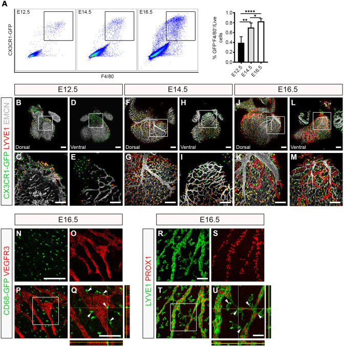
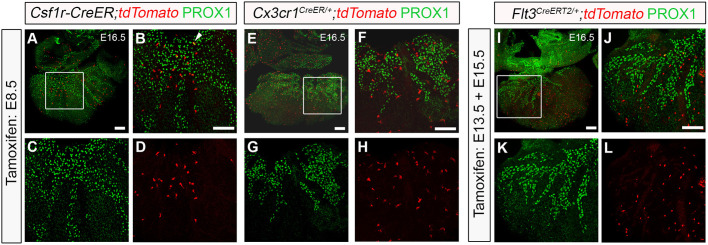
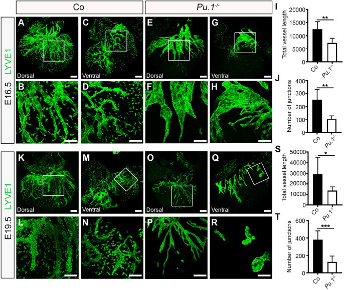
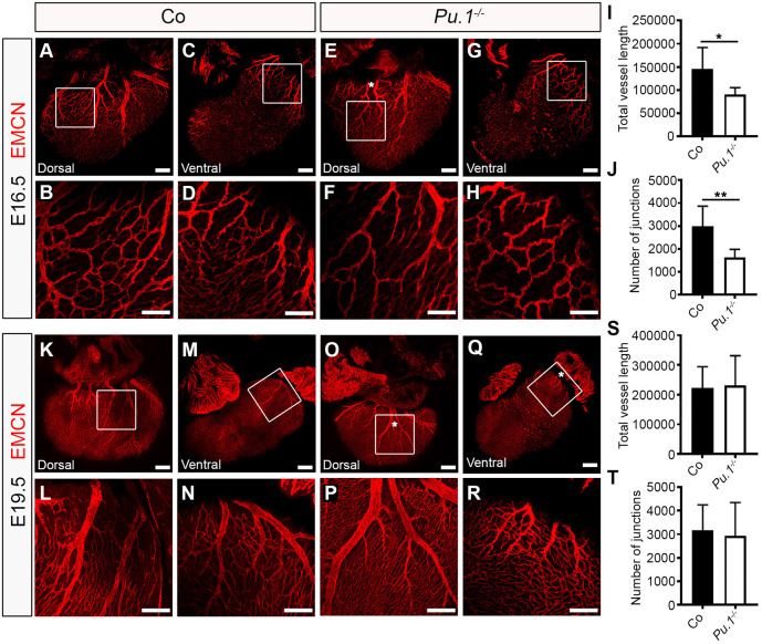
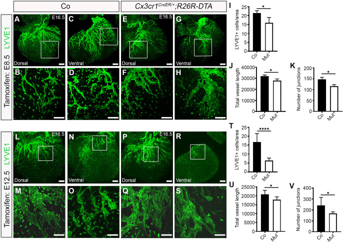
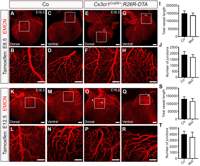
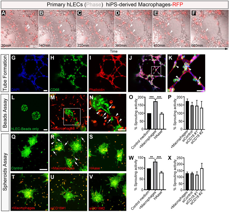
Macrophages are components of the innate immune system with key roles in tissue inflammation and repair. It is now evident that macrophages also support organogenesis, but few studies have characterized their identity, ontogeny and function during heart development. Here, we show that the distribution and prevalence of resident macrophages in the subepicardial compartment of the developing heart coincides with the emergence of new lymphatics, and that macrophages interact closely with the nascent lymphatic capillaries. Consequently, global macrophage deficiency led to extensive vessel disruption, with mutant hearts exhibiting shortened and mis-patterned lymphatics. The origin of cardiac macrophages was linked to the yolk sac and foetal liver. Moreover, the Cx3cr1+ myeloid lineage was found to play essential functions in the remodelling of the lymphatic endothelium. Mechanistically, macrophage hyaluronan was required for lymphatic sprouting by mediating direct macrophage-lymphatic endothelial cell interactions. Together, these findings reveal insight into the role of macrophages as indispensable mediators of lymphatic growth during the development of the mammalian cardiac vasculature.

Keywords: Cardiac lymphatics; Cell adhesion; Coronaries; Hyaluronan; Macrophages; Vessel growth and patterning.


Conflict of interest statement

Competing interestsP.R.R. is co-founder and equity holder in OxStem Cardio, an Oxford University spin-out that seeks to exploit therapeutic strategies stimulating endogenous repair in cardiovascular regenerative medicine.

Figures

Fig. 1.
Fig. 1.
Tissue-resident macrophages are closely associated with the developing cardiac lymphatics. (A) Representative histograms and percentage of tissue-resident macrophages over total number of live singlet cells in the developing heart at embryonic days (E) 12.5, E14.5 and E16.5 measured by flow cytometry for GFP and F4/80 in Cx3cr1GFP/+ reporter embryos. GFP+F4/80+ cells are defined as macrophages. Data are mean±s.e.m.; n=6 hearts per group from at least three independent litters. Significant differences (P-values) were calculated using one-way ANOVA followed by Tukey's multiple comparison test (*P≤0.05; **P≤0.01; ****P≤0.0001). (B-M) Whole-mount immunostaining for GFP (green), LYVE1 (red) and EMCN (white) to visualize the sub-epicardial tissue-resident macrophages, coronary vessels (capillaries and veins) and lymphatic plexus (respectively) in both the dorsal and ventral aspects of hearts derived from Cx3cr1GFP/+ embryos at E12.5 (B-E), E14.5 (F-I) and E16.5 (J-M). (C,E,G,I,K,M) Magnified views of boxes shown in B,D,F,H,J,L. LYVE1 reactivity is detected in the lymphatic endothelium and tissue-resident macrophages. (N-Q) GFP (green) and VEGFR3 (red) immunostaining of whole-mount hearts derived from CD68-GFP-expressing embryos at E16.5. (Q) Orthogonal view of the box shown in P; white arrowheads indicate close association of CD68-GFP+ macrophages with VEGFR3-expressing lymphatic vessels. (R-U) LYVE1 (green) and PROX1 (red) immunostaining of whole-mount hearts derived from C57BL6 embryos at E16.5. (U) Orthogonal view of the box shown in T; white arrowheads indicate LYVE1+ macrophages interacting with fusing lymphatic tip cells labelled with PROX1 (nuclear) and LYVE1 (membrane). n=3-6 hearts per group from at least three independent litters. Scale bars: 100 µm in B-N; 25 µm in Q; 200 µm in R; 50 µm in U.
Fig. 2.
Fig. 2.
Yolk sac-derived Csf1r+ and Cx3cr1+ lineages are associated with cardiac lymphatic growth and expansion. (A-D) Genetic lineage-tracing based on the activity of the Csf1r-CreER;tdTomato transgene induced by tamoxifen administration at embryonic day (E) 8.5. Whole hearts were analysed for Tomato (red) and PROX1 (green) expression at E16.5. (B-D) Magnified views of the box shown in A; white arrowhead indicates colocalization of native Tomato and PROX1 immunoreactivity. (E-H) Genetic lineage-tracing based on the activity of the Cx3cr1CreER/+;tdTomato transgene induced by tamoxifen administration at E8.5. Whole hearts were analyzed for Tomato (red) and PROX1 (green) expression at E16.5. (F-H) Magnified views of the box shown in E. (I-L) Genetic lineage-tracing based on the activity of the Flt3CreERT2/+;tdTomato transgene induced by repeated tamoxifen administration at E13.5 and E15.5. Whole hearts were analyzed for Tomato (red) and PROX1 (green) expression at E16.5. (J-L) Magnified views of the box shown in I. n=3-6 hearts per group from at least three independent litters. Scale bars: 100 µm.
Fig. 3.
Fig. 3.
Macrophages are essential for growth and branching of the cardiac lymphatic network. (A-H) Whole-mount immunostaining for LYVE1 (green) to visualize the sub-epicardial lymphatic plexus in both the dorsal and ventral aspects of hearts derived from littermate control (co; A-D) or Pu.1−/− (E-H) embryos at E16.5. (B,D,F,H) Magnified views of boxes shown in A,C,E,G. LYVE1 reactivity is absent in tissue-resident macrophages in Pu.1−/− hearts compared with control littermates, confirming absence of macrophages. (I,J) Quantification of total vessel length (µm; I) and number of lymphatic vessel junctions (J) in control versus Pu.1−/− hearts at E16.5. Data are mean±s.e.m.; n=6 hearts per group from three independent litters. Significant differences (P-values) were calculated using an unpaired, two-tailed Student's t-test (**P≤0.01). (K-R) Whole-mount immunostaining for LYVE1 (green) to visualize the sub-epicardial lymphatic plexus in both the dorsal and ventral aspects of hearts derived from littermate control (co; K-N) or Pu.1−/− (O-R) embryos at E19.5. (L,N,P,R) Magnified views of boxes shown in K,M,O,Q. (S,T) Quantification of total vessel length (µm; S) and number of lymphatic vessel junctions (T) in control versus Pu.1−/− hearts at E19.5. Data are mean±s.e.m.; control, n=5 hearts; Pu.1−/−, n=6 hearts from four independent litters. Significant differences (P-values) were calculated using an unpaired, two-tailed Student's t-test (*P≤0.05; ***P≤0.001). Scale bars: 100 µm.
Fig. 4.
Fig. 4.
Macrophages regulate coronary growth and patterning. (A-H) Whole-mount immunostaining for EMCN (red) to visualize the sub-epicardial coronary vessels (capillaries and veins) in both the dorsal and ventral aspects of hearts derived from littermate control (co; A-D) or Pu.1−/− (E-H) embryos at E16.5. (B,D,F,H) Magnified views of boxes shown in A,C,E,G. White asterisk indicates patterning defects, i.e. an extra mid-branch, of the coronary veins on the dorsal aspect of Pu.1−/− hearts (compared E with A). (I,J) Quantification of total vessel length (µm; I) and number of vessel junctions (J) in control versus Pu.1−/− hearts at E16.5. Data are mean±s.e.m.; n=6 hearts per group from three independent litters. Significant differences (P-values) were calculated using an unpaired, two-tailed Student's t-test (*P≤0.05; **P≤0.01). (K-R) Whole-mount immunostaining for EMCN (red) to visualize the sub-epicardial coronary vessels (capillaries and veins) in both the dorsal and ventral aspects of hearts derived from littermate control (co; K-N) or Pu.1−/− (O-R) embryos at E19.5. (L,N,P,R) Magnified views of boxes shown in K,M,O,Q. White asterisks indicate patterning defects, i.e. extra branches, of the coronary veins on both the dorsal and ventral aspects of Pu.1−/− hearts (compare O,Q with K,M). (S,T) Quantification of total vessel length (µm; S) and number of vessel junctions (T) in control versus Pu.1−/− hearts at E19.5. Data are mean±s.e.m.; control, n=5 hearts; Pu.1−/−, n=6 hearts from four independent litters. No significant differences were determined using an unpaired, two-tailed Student's t-test. Scale bars: 100 µm in A-D,F-H,K-R; 1 mm in E.
Fig. 5.
Fig. 5.
Ablation of yolk sac-derived macrophages impairs cardiac lymphatic growth and branching. (A-H) Whole-mount immunostaining for LYVE1 (green) to visualize the sub-epicardial lymphatic plexus in both the dorsal and ventral aspects of E16.5 hearts-derived from littermate control (co; A-D) or Cx3cr1CreER/+;R26R-DTA (Mut; E-H) embryos that were tamoxifen induced at E8.5. (B,D,F,H) Magnified views of boxes shown in A,C,E,G. (I-K) Quantification of LYVE1+ cells per ventricular surface area (I), total vessel length (µm; J) and number of lymphatic vessel junctions (K) in control versus mutant hearts at E16.5. Data are mean±s.e.m.; n=3 hearts per group from two independent litters. Significant differences (P-values) were calculated using an unpaired, two-tailed Student's t-test (*P≤0.05). (L-S) Whole-mount immunostaining for LYVE1 (green) to visualize the sub-epicardial lymphatic plexus in both the dorsal and ventral aspects of E16.5 hearts derived from littermate control (co; L-O) or Cx3cr1CreER/+;R26R-DTA (Mut; P-S) embryos that were tamoxifen induced at E12.5. (M,O,Q,S) Magnified views of boxes shown in L,N,P,R. (T-V) Quantification of LYVE1+ cells per ventricular surface area (T), total vessel length (µm; U) and number of lymphatic vessel junctions (V) in control versus Mut hearts at E16.5. Data are mean±s.e.m.; control, n=8 hearts; Mut, n=7 hearts from three independent litters. Significant differences (P-values) were calculated using an unpaired, two-tailed Student's t-test (*P≤0.05; ****P≤0.0001). Scale bars: 100 µm.
Fig. 6.
Fig. 6.
Ablation of yolk sac-derived macrophages disrupts coronary vein patterning but does not impact on vessel growth or branching. (A-H) Whole-mount immunostaining for EMCN (red) to visualize the sub-epicardial coronary vessels (capillaries and veins) in both the dorsal and ventral aspects of E16.5 hearts derived from littermate control (co; A-D) or Cx3cr1CreER/+;R26R-DTA (Mut; E-H) embryos that were tamoxifen induced at E8.5. (B,D,F,H) Magnified views of boxes shown in A,C,E,G. White asterisks indicate patterning defects, i.e. an extra branch, of the coronary veins on the dorsal and ventral aspects of Cx3cr1CreER/+;R26R-DTA hearts (compared E,G with A,C), akin to Pu.1-null hearts (compared with Fig. 4). (I,J) Quantification of total vessel length (µm; I) and number of vessel junctions (J) in control versus mutant hearts at E16.5. Data are mean±s.e.m.; n=3 hearts per group from two independent litters. No significant differences were determined using an unpaired, two-tailed Student's t-test. (K-R) Whole-mount immunostaining for EMCN (red) to visualize the sub-epicardial coronary vessels (capillaries and veins) in both the dorsal and ventral aspects of E16.5 hearts derived from littermate control (co; K-N) or Cx3cr1CreER/+;R26R-DTA (Mut; O-R) embryos that were tamoxifen induced at E12.5. (L,N,P,R) Magnified views of boxes shown in K,M,O,Q. White asterisks indicate patterning defects, i.e. an extra branch, of the coronary veins on the dorsal and ventral aspects of Cx3cr1CreER/+;R26R-DTA hearts (compare O,Q with K,M), akin to Pu.1-null hearts (compared with Fig. 4). (S,T) Quantification of total vessel length (µm; S) and number of vessel junctions (T) in control versus mutant hearts at E16.5. Data are mean±s.e.m.; control, n=8 hearts; mutant, n=7 hearts from three independent litters. No significant differences were determined using an unpaired, two-tailed Student's t-test. Scale bars: 100 µm.
Fig. 7.
Fig. 7.
Macrophages promote lymphatic cell plexus formation and sprouting. (A-F) Representative frames of time-lapse experiments using primary human lymphatic endothelial cells (hLECs; phase-contrast) co-cultured with human iPS-derived macrophages labelled with RFP (red), showing close association between macrophages and LECs, and a change in macrophage cell morphology (white arrowheads) contacting the expanding lymphatic plexus to facilitate LEC tube formation. (G-K) DAPI (blue), CD68 (green) and phalloidin (red) staining of primary hLEC and hiPSC-macrophage co-cultures. (K) Magnified view of box shown in J. Arrowheads indicate macrophages. (L-N) Representative staining of lymphatic capillary sprouting from beads coated with primary hLECs only (green; control medium; L) or co-cultured with hiPSC-derived macrophages-RFP (red; +macrophages; M). (N) Magnified view of the box shown in M. Arrows and arrowheads indicate lymphatic sprouts and macrophages, respectively. (O,P) Quantification of the percentage sprouting activity defined as (number capillary sprouts/number of beads)×100. Data are normalized against the control group. Data are mean±s.e.m.; control, n=6; +macrophages, n=6; HAase 30 U/ml, n=3; siControl, n=3; siCD18 #1, n=3; siCD18 #2, n=3 independent experiments, with three replicates/experiment. Significant differences (P-values) were calculated using one-way ANOVA followed by Tukey's multiple comparison test (***P≤0.001). (Q-V) Representative staining of lymphatic capillary sprouting from hLECs aggregates/spheroids only (green; control medium; Q) or co-cultured with hiPSC-derived macrophages-RFP (red; +macrophages; R,T); and of hiPSC-derived macrophages-RFP pre-treated with HAase 30 U/ml (S) or co-cultured with hiPSC-derived macrophages-RFP pre-treated with siRNA oligonucleotides against ITGB2/CD18 (siCD18 #1, U; siCD18 #2, V). (W,X) Quantification of the percentage of sprouting activity defined as (number capillary sprouts/number of beads)×100. Data were normalized against the control group. Data are mean±s.e.m.; control, n=5; +macrophages, n=4; HAase 30 U/ml, n=4; siControl, n=3; siCD18 #1, n=3; siCD18 #2, n=3 independent experiments, with three replicates/experiment. Significant differences (P-values) were calculated using one-way ANOVA followed by Tukey's multiple comparison test (**P≤0.01; ***P≤0.001). Scale bars: 100 µm in A-M,R-V; 50 µm in N; 250 µm in Q.

Comment in

References

    1. Aurora, A. B., Porrello, E. R., Tan, W., Mahmoud, A. I., Hill, J. A., Bassel-Duby, R., Sadek, H. A. and Olson, E. N. (2014). Macrophages are required for neonatal heart regeneration. J. Clin. Invest. 124, 1382-1392. 10.1172/JCI72181 - DOI - PMC - PubMed
    1. Becht, E., McInnes, L., Healy, J., Dutertre, C.-A., Kwok, I. W. H., Ng, L. G., Ginhoux, F. and Newell, E. W. (2018). Dimensionality reduction for visualizing single-cell data using UMAP. Nat. Biotechnol. 37, 38-44. 10.1038/nbt.4314 - DOI - PubMed
    1. Benz, C., Martins, V. C., Radtke, F. and Bleul, C. C. (2008). The stream of precursors that colonizes the thymus proceeds selectively through the early T lineage precursor stage of T cell development. J. Exp. Med. 205, 1187-1199. 10.1084/jem.20072168 - DOI - PMC - PubMed
    1. Boyer, S. W., Schroeder, A. V., Smith-Berdan, S. and Forsberg, E. C. (2011). All hematopoietic cells develop from hematopoietic stem cells through Flk2/Flt3-positive progenitor cells. Cell Stem Cell 9, 64-73. 10.1016/j.stem.2011.04.021 - DOI - PMC - PubMed
    1. Brachtendorf, G., Kuhn, A., Samulowitz, U., Knorr, R., Gustafsson, E., Potocnik, A. J., Fässler, R. and Vestweber, D. (2001). Early expression of endomucin on endothelium of the mouse embryo and on putative hematopoietic clusters in the dorsal aorta. Dev. Dyn. 222, 410-419. 10.1002/dvdy.1199 - DOI - PubMed

Publication types

Substances

LinkOut - more resources