. 2021 Jan 18;11(1):1702.
doi: 10.1038/s41598-020-80573-x.

Predicting mammalian species at risk of being infected by SARS-CoV-2 from an ACE2 perspective

Affiliations

Predicting mammalian species at risk of being infected by SARS-CoV-2 from an ACE2 perspective

Yulong Wei et al. Sci Rep. .

Erratum in

Abstract

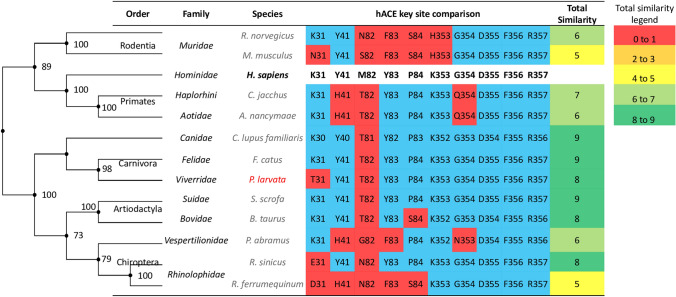
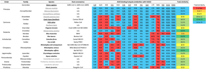
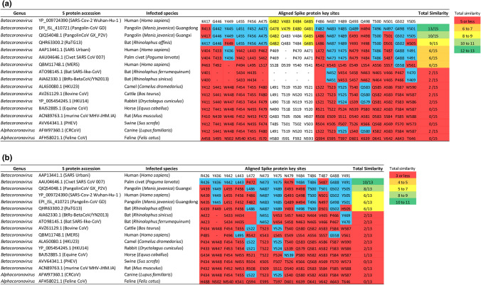
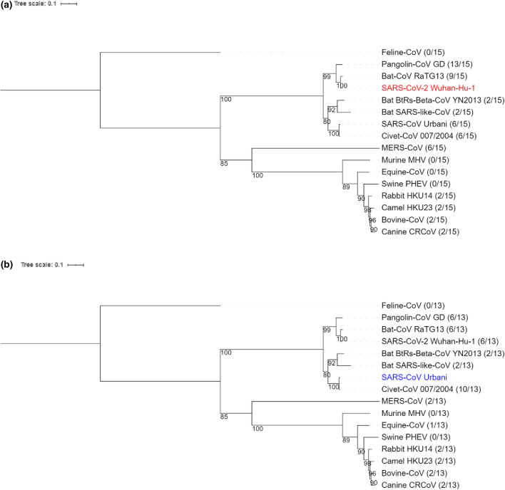
SARS-CoV-2 can transmit efficiently in humans, but it is less clear which other mammals are at risk of being infected. SARS-CoV-2 encodes a Spike (S) protein that binds to human ACE2 receptor to mediate cell entry. A species with a human-like ACE2 receptor could therefore be at risk of being infected by SARS-CoV-2. We compared between 132 mammalian ACE2 genes and between 17 coronavirus S proteins. We showed that while global similarities reflected by whole ACE2 gene alignments are poor predictors of high-risk mammals, local similarities at key S protein-binding sites highlight several high-risk mammals that share good ACE2 homology with human. Bats are likely reservoirs of SARS-CoV-2, but there are other high-risk mammals that share better ACE2 homologies with human. Both SARS-CoV-2 and SARS-CoV are closely related to bat coronavirus. Yet, among host-specific coronaviruses infecting high-risk mammals, key ACE2-binding sites on S proteins share highest similarities between SARS-CoV-2 and Pangolin-CoV and between SARS-CoV and Civet-CoV. These results suggest that direct coronavirus transmission from bat to human is unlikely, and that rapid adaptation of a bat SARS-like coronavirus in different high-risk intermediate hosts could have allowed it to acquire distinct high binding potential between S protein and human-like ACE2 receptors.


Conflict of interest statement

The authors declare no competing interests.

Figures

Figure 1
Figure 1
A sample comparison between mammalian ACE2 genes. Global similarities represented by whole ACE2 gene comparisons (the phylogenetic tree on the left) poorly predict mammals at high risk of being infected by SARS-CoV; whereas local similarities at key human ACE2 sites (the table on the right) show that species in the Carnivora, Artiodactyla and Chiroptera orders are at high risk of SARS-CoV infection. The tips of the phylogenetic tree are aligned with the corresponding species listed in the table. Species in red is the masked palm civet, a possible intermediate host of SARS-CoV. Species in black bold is human, and all other species are in grey. Amino acids highlighted in blue and red are matching and mis-matching sites, respectively, between mammalian ACE2 and human ACE2 at key SARS-CoV S protein binding sites. Total similarity in the table is calculated as the sum of matching amino acids between mammalian and human ACE2 at key binding sites. A comparison of key ACE2 sites involved in SARS-CoV binding between human and 131 mammals is shown in Supplementary Figure S3.
Figure 2
Figure 2
Comparisons at key ACE2 sites between human and a sample of mammals belong to ten orders. Species that have been experimentally identified as susceptible to SARS-CoV-2 infection are underlined. Species that can be infected by their own host-specific coronaviruses are in black bold, and other species are in grey. Highlighted blue and red are matching and mis-matching amino acid residues, respectively, between mammalian ACE2 and human ACE2 at key SARS-CoV-2 S protein binding sites. Underlined amino acids are conserved among species known to be infected by SARS-CoV-2. Total similarity designates the total number of matching amino acid residues with respect to human ACE2. The total similarity score for mammalian ACE2 in blue highlights perfect site similarities (14–15 matching sites), in green highlights high similarity (12–13 matching sites), in light green highlights medium–high similarity (10–11 matching sites), in yellow highlights medium similarity (8–9 matching sites), in orange highlights medium–low similarity (6–7 matching sites), and in red highlights low similarity (5 or less matching sites). A comparison of key ACE2 sites involved in SARS-CoV-2 binding between human and 131 mammals is shown in Supplementary Figure S1.
Figure 3
Figure 3
Amino acid comparisons at key S protein sites among 17 mammalian-specific coronaviruses. (a) Displays the conservation of key ACE2-binding sites on SARS-CoV-2 S protein by 16 mammalian-specific S proteins. (b) Displays the conservation of key ACE2-binding sites on SARS-CoV S protein by 16 mammalian-specific S proteins. Highlighted blue and red are matching and mis-matching amino acid residues, respectively, between mammalian coronaviruses and the two human SARS coronaviruses at key S protein sites. Highlighted yellow is the 482–485 GVEG motif found in SARS-CoV-2. Total similarity designates the total number of matching amino acid residues with respect to SARS-CoV-2 or SARS-CoV, and scores in green highlights high similarity, in light green highlights medium–high similarity, in yellow highlights medium similarity, in orange highlights medium–low similarity, and in red highlights low similarity.
Figure 4
Figure 4
Phylogenetic reconstruction of 16 MAFFT G-INS-i aligned coronavirus S protein-coding genes. Infected mammalian species belong to the Artiodactyla, Canivora, Chiroptera, Lagomorpha, Perissodactyla, Primates, Pholidota and Rodentia orders. The phylogenetic tree is constructed using the maximum-likelihood-based PHYML approach, with best model = WAG + G + I + F, and Bootstrap = 100. Pangolin-CoV GX strain was excluded in favor of Pangolin-CoV GD strain having better sequence coverage. The ratio appended to each coronavirus indicates the total number of amino acid matches when coronavirus S protein gene was compared to (a) SARS-CoV-2 S protein gene (in red) at its 15 key ACE2-binding sites and to (b) SARS-CoV S protein gene (in blue) at its 13 key ACE-2 binding sites.

References

    1. Cui J, Li F, Shi ZL. Origin and evolution of pathogenic coronaviruses. Nat. Rev. Microbiol. 2019;17:181–192. doi: 10.1038/s41579-018-0118-9. - DOI - PMC - PubMed
    1. Lan J, et al. Structure of the SARS-CoV-2 spike receptor-binding domain bound to the ACE2 receptor. Nature. 2020;581:215–220. doi: 10.1038/s41586-020-2180-5. - DOI - PubMed
    1. Li F, Li W, Farzan M, Harrison SC. Structure of SARS coronavirus spike receptor-binding domain complexed with receptor. Science. 2005;309:1864–1868. doi: 10.1126/science.1116480. - DOI - PubMed
    1. Li W, et al. Receptor and viral determinants of SARS-coronavirus adaptation to human ACE2. Embo. J. 2005;24:1634–1643. doi: 10.1038/sj.emboj.7600640. - DOI - PMC - PubMed
    1. Shang J, et al. Structural basis of receptor recognition by SARS-CoV-2. Nature. 2020;30:020–2179. - PMC - PubMed

Publication types

MeSH terms

Substances

LinkOut - more resources