

Perinatal SSRI exposure affects brain functional activity associated with whisker stimulation in adolescent and adult rats
- PMID: 33462357
- PMCID: PMC7814075
- DOI: 10.1038/s41598-021-81327-z
Perinatal SSRI exposure affects brain functional activity associated with whisker stimulation in adolescent and adult rats
Abstract
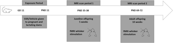
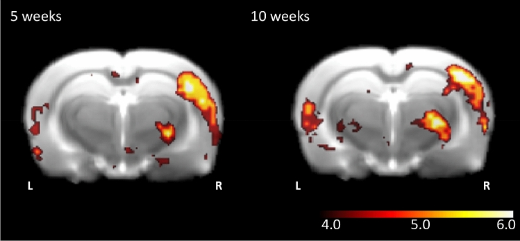
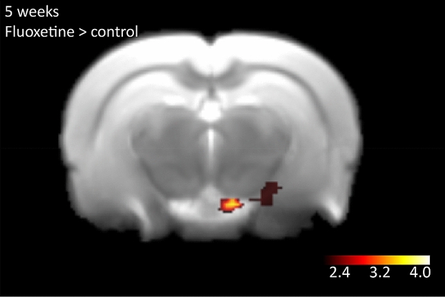
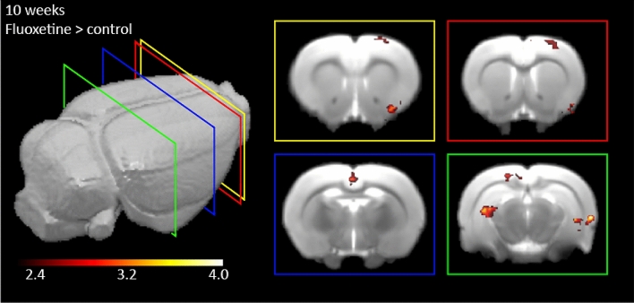
Selective serotonin reuptake inhibitors (SSRI), such as fluoxetine, are used as first-line antidepressant medication during pregnancy. Since SSRIs cross the placenta the unborn child is exposed to the maternal SSRI medication, resulting in, amongst others, increased risk for autism in offspring. This likely results from developmental changes in brain function. Studies employing rats lacking the serotonin transporter have shown that elevations in serotonin levels particularly affect the development of the whisker related part of the primary somatosensory (barrel) cortex. Therefore, we hypothesized that serotonin level disturbances during development alter brain activity related to whisker stimulation. We treated female dams with fluoxetine or vehicle from gestational day 11 onwards for 21 days. We investigated offspring's brain activity during whisker stimulation using functional magnetic resonance imaging (fMRI) at adolescence and adulthood. Our results indicate that adolescent offspring displayed increased activity in hippocampal subareas and the mammillary body in the thalamus. Adult offspring exhibited increased functional activation of areas associated with (higher) sensory processing and memory such as the hippocampus, perirhinal and entorhinal cortex, retrospinal granular cortex, piriform cortex and secondary visual cortex. Our data imply that perinatal SSRI exposure leads to complex alterations in brain networks involved in sensory perception and processing.
Conflict of interest statement
The authors declare no competing interests.
Figures
Similar articles
-
Gestational stress and fluoxetine treatment differentially affect plasticity, methylation and serotonin levels in the PFC and hippocampus of rat dams.Neuroscience. 2016 Jul 7;327:32-43. doi: 10.1016/j.neuroscience.2016.03.068. Epub 2016 Apr 11. Neuroscience. 2016. PMID: 27060483
-
Developmental fluoxetine exposure and prenatal stress alter sexual differentiation of the brain and reproductive behavior in male rat offspring.Psychoneuroendocrinology. 2013 Sep;38(9):1618-29. doi: 10.1016/j.psyneuen.2013.01.007. Epub 2013 Feb 8. Psychoneuroendocrinology. 2013. PMID: 23399049
-
Perinatal fluoxetine dose-dependently affects prenatal stress-induced neurobehavioural abnormalities, HPA-axis functioning and underlying brain alterations in rat dams and their offspring.Reprod Toxicol. 2021 Sep;104:27-43. doi: 10.1016/j.reprotox.2021.06.014. Epub 2021 Jun 26. Reprod Toxicol. 2021. PMID: 34186199
-
Of rodents and humans: A comparative review of the neurobehavioral effects of early life SSRI exposure in preclinical and clinical research.Int J Dev Neurosci. 2016 Jun;51:50-72. doi: 10.1016/j.ijdevneu.2016.04.008. Epub 2016 May 7. Int J Dev Neurosci. 2016. PMID: 27165448 Free PMC article. Review.
-
The effects of maternal antidepressant use on offspring behaviour and brain development: Implications for risk of neurodevelopmental disorders.Neurosci Biobehav Rev. 2017 Sep;80:743-765. doi: 10.1016/j.neubiorev.2017.06.008. Epub 2017 Jun 16. Neurosci Biobehav Rev. 2017. PMID: 28629713 Review.
Cited by
-
High-resolution awake mouse fMRI at 14 tesla.Elife. 2025 Jan 9;13:RP95528. doi: 10.7554/eLife.95528. Elife. 2025. PMID: 39786364 Free PMC article.
-
High-resolution awake mouse fMRI at 14 Tesla.bioRxiv [Preprint]. 2024 Sep 23:2023.12.08.570803. doi: 10.1101/2023.12.08.570803. bioRxiv. 2024. Update in: Elife. 2025 Jan 09;13:RP95528. doi: 10.7554/eLife.95528. PMID: 38106227 Free PMC article. Updated. Preprint.
-
Transcriptional changes in specific subsets of Drosophila neurons following inhibition of the serotonin transporter.Transl Psychiatry. 2023 Jun 24;13(1):226. doi: 10.1038/s41398-023-02521-3. Transl Psychiatry. 2023. PMID: 37355701 Free PMC article.
-
Chirality of antidepressive drugs: an overview of stereoselectivity.Asian Biomed (Res Rev News). 2022 Apr 29;16(2):55-69. doi: 10.2478/abm-2022-0008. eCollection 2022 Apr. Asian Biomed (Res Rev News). 2022. PMID: 37551287 Free PMC article. Review.
-
Transcriptional changes in specific subsets of Drosophila neurons following inhibition of the serotonin transporter.Res Sq [Preprint]. 2023 Mar 17:rs.3.rs-2626506. doi: 10.21203/rs.3.rs-2626506/v1. Res Sq. 2023. Update in: Transl Psychiatry. 2023 Jun 24;13(1):226. doi: 10.1038/s41398-023-02521-3. PMID: 36993644 Free PMC article. Updated. Preprint.
References
-
- Organization, G. W. H. Depression and other common mental disorders: Global health estimates. License: CC BY-NC-SA 3.0 IGO (2017).
-
- Verweij, G. & Houben-van Herten, M. Depressiviteit en antidepressiva in Nederland. Centraal Bureau voor de Statistiek (2013).
Publication types
MeSH terms
Substances
LinkOut - more resources
Full Text Sources
Other Literature Sources
