The evolving cardiac lymphatic vasculature in development, repair and regeneration
- PMID: 33462421
- PMCID: PMC7812989
- DOI: 10.1038/s41569-020-00489-x
The evolving cardiac lymphatic vasculature in development, repair and regeneration
Abstract
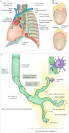
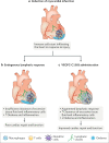
The lymphatic vasculature has an essential role in maintaining normal fluid balance in tissues and modulating the inflammatory response to injury or pathogens. Disruption of normal development or function of lymphatic vessels can have severe consequences. In the heart, reduced lymphatic function can lead to myocardial oedema and persistent inflammation. Macrophages, which are phagocytic cells of the innate immune system, contribute to cardiac development and to fibrotic repair and regeneration of cardiac tissue after myocardial infarction. In this Review, we discuss the cardiac lymphatic vasculature with a focus on developments over the past 5 years arising from the study of mammalian and zebrafish model organisms. In addition, we examine the interplay between the cardiac lymphatics and macrophages during fibrotic repair and regeneration after myocardial infarction. Finally, we discuss the therapeutic potential of targeting the cardiac lymphatic network to regulate immune cell content and alleviate inflammation in patients with ischaemic heart disease.
Conflict of interest statement
P.R.R. is co-founder and equity holder in OxStem Cardio, an Oxford University spin-out that seeks to exploit therapeutic strategies stimulating endogenous repair in cardiovascular regenerative medicine. The other authors declare no competing interests.
Figures


Similar articles
-
Distinct origins and molecular mechanisms contribute to lymphatic formation during cardiac growth and regeneration.Elife. 2019 Nov 8;8:e44153. doi: 10.7554/eLife.44153. Elife. 2019. PMID: 31702554 Free PMC article.
-
Anatomy and development of the cardiac lymphatic vasculature: Its role in injury and disease.Clin Anat. 2016 Apr;29(3):305-15. doi: 10.1002/ca.22638. Epub 2015 Nov 23. Clin Anat. 2016. PMID: 26443964 Review.
-
[Cardiac lymphatic system].Med Sci (Paris). 2017 Aug-Sep;33(8-9):765-770. doi: 10.1051/medsci/20173308022. Epub 2017 Sep 18. Med Sci (Paris). 2017. PMID: 28945567 Review. French.
-
Formation and Growth of Cardiac Lymphatics during Embryonic Development, Heart Regeneration, and Disease.Cold Spring Harb Perspect Biol. 2020 Jun 1;12(6):a037176. doi: 10.1101/cshperspect.a037176. Cold Spring Harb Perspect Biol. 2020. PMID: 31818858 Free PMC article. Review.
-
Cardiac lymphatics in health and disease.Nat Rev Cardiol. 2019 Jan;16(1):56-68. doi: 10.1038/s41569-018-0087-8. Nat Rev Cardiol. 2019. PMID: 30333526 Review.
Cited by
-
The Enigmas of Lymphatic Muscle Cells: Where Do They Come From, How Are They Maintained, and Can They Regenerate?Curr Rheumatol Rev. 2023 Jun 5;19(3):246-259. doi: 10.2174/1573397119666230127144711. Curr Rheumatol Rev. 2023. PMID: 36705238 Free PMC article. Review.
-
Advancing 3D Engineered In Vitro Models for Heart Failure Research: Key Features and Considerations.Bioengineering (Basel). 2024 Dec 3;11(12):1220. doi: 10.3390/bioengineering11121220. Bioengineering (Basel). 2024. PMID: 39768038 Free PMC article. Review.
-
Progress of Mitochondrial Function Regulation in Cardiac Regeneration.J Cardiovasc Transl Res. 2024 Oct;17(5):1097-1105. doi: 10.1007/s12265-024-10514-w. Epub 2024 Apr 22. J Cardiovasc Transl Res. 2024. PMID: 38647881 Review.
-
The Zebrafish Cardiac Endothelial Cell-Roles in Development and Regeneration.J Cardiovasc Dev Dis. 2021 May 1;8(5):49. doi: 10.3390/jcdd8050049. J Cardiovasc Dev Dis. 2021. PMID: 34062899 Free PMC article. Review.
-
A cigarette filter-derived biomimetic cardiac niche for myocardial infarction repair.Bioact Mater. 2024 Feb 14;35:362-381. doi: 10.1016/j.bioactmat.2024.02.012. eCollection 2024 May. Bioact Mater. 2024. PMID: 38379697 Free PMC article.
References
Publication types
MeSH terms
Grants and funding
LinkOut - more resources
Full Text Sources
Other Literature Sources
Medical
Miscellaneous

