Review
. 2021 Mar;20(3):217-243.
doi: 10.1038/s41573-020-00093-1. Epub 2021 Jan 18.

The clinical impact of glycobiology: targeting selectins, Siglecs and mammalian glycans

Affiliations
Review

The clinical impact of glycobiology: targeting selectins, Siglecs and mammalian glycans

Benjamin A H Smith et al. Nat Rev Drug Discov. 2021 Mar.

Erratum in

Abstract

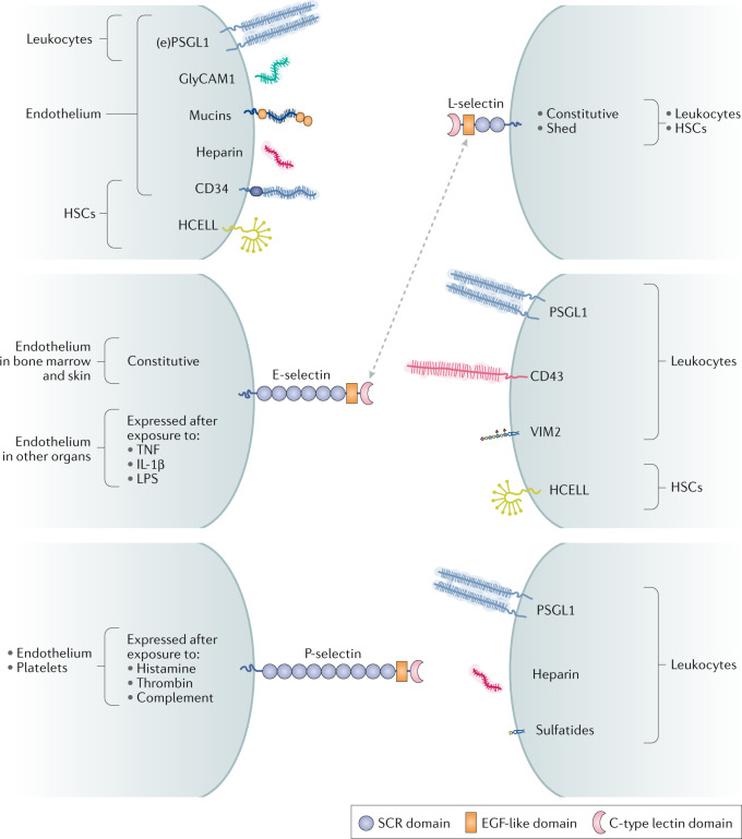
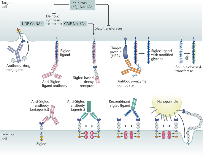
Carbohydrates - namely glycans - decorate every cell in the human body and most secreted proteins. Advances in genomics, glycoproteomics and tools from chemical biology have made glycobiology more tractable and understandable. Dysregulated glycosylation plays a major role in disease processes from immune evasion to cognition, sparking research that aims to target glycans for therapeutic benefit. The field is now poised for a boom in drug development. As a harbinger of this activity, glycobiology has already produced several drugs that have improved human health or are currently being translated to the clinic. Focusing on three areas - selectins, Siglecs and glycan-targeted antibodies - this Review aims to tell the stories behind therapies inspired by glycans and to outline how the lessons learned from these approaches are paving the way for future glycobiology-focused therapeutics.


Conflict of interest statement

C.R.B. is a co-founder of Redwood Bioscience, Enable Biosciences, Palleon Pharmaceuticals, InterVenn Bio, Lycia Therapeutics and OliLux Biosciences, and a member of the Board of Directors of Eli Lilly. B.A.H.S. is a shareholder of GlycoMimetics.

Figures

Fig. 1
Fig. 1. Selectins and their primary ligands.
In humans, selectin and selectin ligands expressed on the endothelium, platelets and other cells (shown on the left) interact with selectin and selectin ligands on leukocytes or haematopoietic stem cells (HSCs) (shown on the right). The ligands for each selectin comprise glycoproteins bearing sialofucosylated glycans that are closely related to sialyl Lewisx (sLex), and, in some cases, glycolipids such as VIM2 and glycosaminoglycans such as heparin. L-selectin is both constitutively expressed and shed from leukocyte cell surfaces. E-selectin and P-selectin are displayed on cells in response to inflammatory stimuli. Mucins refers to endothelial glycoproteins not otherwise depicted that function as L-selectin ligands, including mucosal addressin cell adhesion molecule 1 (MAdCAM1), podocalyxin-like protein, Sgp200, endoglycan and endomucin. E-selectin is constitutively expressed on the endothelium in the bone marrow and skin, but requires exposure to inflammatory cytokines to be expressed in other organs. The dotted arrow indicates that sLex on L-selectin itself is a ligand for E-selectin. EGF, epidermal growth factor; (e)PSGL1, (endothelial) P-selectin glycoprotein ligand 1; GlyCAM1, glycosylation-dependent cell adhesion molecule 1; HCELL, haematopoietic cell E-/L-selectin ligand; IL-1β, interleukin-1β; LPS, lipopolysaccharide; SCR, short consensus repeat (Sushi domain); TNF, tumour necrosis factor.
Fig. 2
Fig. 2. P-selectin engages both glycan and protein portions of PSGL1.
P-selectin forms a complex with P-selectin glycoprotein ligand 1 (PSGL1). a | Schematic depicting the polar contacts (dashed lines) between SGP-3, a sulfoglycopeptide derived from the amino terminus of PSGL1 (shown in the red box using single-letter amino acid codes), and P-selectin (shown in the black box using three-letter amino acid codes). The glycan attached to Thr16 is represented using the colour-coded symbol nomenclature for glycans (SNFG). Note that sulfotyrosine 5 (Tys5) was poorly resolved in the crystal structure and is therefore depicted without polar contacts. b | Crystal structure of P-selectin bound to SGP-3 (Protein Data Bank identifier: 1G1S). P-selectin has two binding surfaces: one interacts with sialyl Lewisx (sLex) and the other interacts with portions of the PSGL1 protein backbone. c | The PSGL1 protein backbone (yellow) makes key contacts with P-selectin (grey) through Pro14 and two sulfotyrosines, Tys7 and Tys10. The third sulfotyrosine that is important for the interaction, Tys5, was modelled as an alanine in the crystal structure. Fuc, fucose; Gal, galactose; GalNAc, N-acetylgalactosamine; GlcNAc, N-acetylglucosamine; Neu5Ac, N-acetylneuraminic acid.
Fig. 3
Fig. 3. Small-molecule selectin inhibitors.
The chemical structures of sialyl Lewisx (sLex) and sialyl Lewisa (sLea) are presented at the top for reference. Structural motifs within the small-molecule selectin inhibitors that have homology to the sialyl Lewis scaffolds are colour coded. The orange highlighted pharmacophore in rivipansel mimics the sulfotyrosines in P-selectin glycoprotein ligand 1 (PSGL1) that are important for the interaction with P-selectin. In sevuparin, the blue and grey shading highlights 2-N-sulfo-6-O-sulfo-glucosamine and iduronic-2-O-sulfate, respectively, which mimic portions of heparan sulfate. In OJ-R9188, replacement of the six-membered fucose ring with fucufuranose maintained binding to E-selectin while increasing resistance to hydrolytic enzymes. Fuc, fucose; Gal, galactose; GlcNAc, N-acetylglucosamine; Neu5Ac, N-acetylneuraminic acid.
Fig. 4
Fig. 4. Siglecs and downstream signalling.
a,b | Protein domains of all members of the human (part a) and mouse (part b) Siglec families. The Siglecs can be broadly divided into the conserved Siglecs (sialoadhesin, CD22, myelin associated glycoprotein (MAG) and Siglec-15) and the CD33-related (CD33r) Siglecs that have diverged more recently on the evolutionary timescale. Cell types that express each Siglec are indicated. Double-headed arrows show functional orthologues among CD33r Siglecs. Murine Siglec-E is considered the functional orthologue of human Siglec-5, Siglec-7 and Siglec-9. Siglec-F is the functional paralogue of human Siglec-8, although it is an orthologue of human Siglec-5 and Siglec-6 (dashed line). Siglec-G is the functional orthologue of human Siglec-10. Human Siglec-XII lacks the arginine essential for sialic acid binding and is non-functional. Chimpanzee Siglec-13 was deleted in humans. Signalling domains in the cytoplasmic tails of each protein are depicted as coloured boxes. The immunoreceptor tyrosine-based inhibitory motif (ITIM) sequence is [I/L/V]xYxx[L/V], the ITIM-like sequence is [D/E]YxE[V/I][R/K], the immunoreceptor tyrosine-based switch motif (ITSM) sequence is TxYxx[V/I], the growth factor receptor-bound protein 2 (GRB2) SH2 binding motif is YxNx and the FYN kinase site is RxxS. Other non-consensus motif tyrosines, such as in murine CD33 and Siglec-F, are not depicted. The GRB2-binding motif in Siglec-10 and Siglec-G is contained within an ITIM. Siglec expression patterns are indicated according to independent reports in the literature. Recent data also suggest that murine T cells express Siglec-E and Siglec-G, and that murine platelets express Siglec-E. c | Domain organization and signalling motifs of other immune cell receptors with known roles in immune modulation, illustrated for comparison. d | Siglecs with ITIMs and ITIM-like signalling motifs may be phosphorylated by SRC family kinases, thereby enabling the recruitment of the protein phosphatases SRC homology region 2 domain-containing phosphatase 1 (SHP1) and SHP2. The ITIM domains in CD33 and Siglec-7 have also been shown to recruit suppressor of cytokine signaling 3 (SOCS3). Siglecs with basic residues in their transmembrane domain enable interactions with the scaffold protein DNAX-activation protein 12 (DAP12). DAP12 contains four immunoreceptor tyrosine-based activation motif (ITAM) domains that, when phosphorylated by SRC family kinases, lead to SYK activation. Figure inspired by ref.. DC, dendritic cell; MyPro, myeloid progenitor; NK cell, natural killer cell; ODC, oligodendrocyte; P, phosphate; pDC, plasmacytoid dendritic cell; SIRPα, signal regulatory protein-α.
Fig. 5
Fig. 5. Modalities for Siglec-targeted therapies.
Antibody–drug conjugates target the Siglecs (CD22 and CD33) that are expressed by cancers, including acute myeloid leukaemia and B cell lymphomas. Anti-Siglec antibodies can either agonize or antagonize Siglec activity. Siglec-blocking antibodies can function as antagonists by preventing ligand binding, but many serve as mild agonists by promoting dimerization. Siglec agonist antibodies can dampen immune cell activity and promote apoptosis. Siglec ligand-blocking antibodies antagonize Siglec function via competitive binding to the Siglec ligand. Recombinant Siglec ligands, shown here as a recombinant glycoprotein fused to an antibody Fc domain, could theoretically act as either receptor agonists or antagonists, likely depending on their ability to cluster Siglecs. Siglec-based decoy receptors comprising a soluble Siglec protein bind to and block the set of ligands for any given Siglec. Therefore, they may antagonize Siglec activity when ligand identities are unclear or diverse. Antibody–enzyme conjugates comprise antibodies directed to target cell-specific antigens conjugated to a glycocalyx editing enzyme such as a sialidase. In the case of an antibody–sialidase conjugate, removal of sialic acid destroys Siglec ligands, thereby antagonizing immune cell Siglecs. Glycosyltransferases in circulation or administered therapeutically may use nucleotide sugars released from platelets to alter the glycocalyx by creating or destroying Siglec binding sites. Nanoparticles such as liposomes bearing Siglec ligands may agonize Siglecs via aggregation or serve as a means for targeted payload delivery. Finally, small-molecule inhibitors, such as the fluorinated sialic acid analogue 3Fax-Neu5Ac, of de novo sialic acid synthesis, or the sialyltransferases, the enzymes responsible for linking sialic acid to nascent glycoproteins and glycolipids, may reduce the sialic acid content of the glycocalyx and destroy Siglec ligands. CMP-Neu5Ac, cytidine monophosphate-N-acetyl-neuraminic acid; HER2, human epidermal growth factor receptor 2 (also known as ERBB2); P, phosphate; UDP-GalNAc, uridine diphosphate-N-acetyl-galactosamine.
Fig. 6
Fig. 6. Tumour-associated carbohydrate antigen vaccines.
Vaccines targeting tumour-associated carbohydrates have evolved from unimolecular vaccines comprising single glycans conjugated to a carrier protein (usually keyhole limpet haemocyanin (KLH)) into more complex multivalent vaccines in which multiple glycans are linked together on a single scaffold. Although glycolipids have been popular targets, some vaccines target mucin-associated glycans. Pharmacophores representing key glycans in each vaccine are shaded in colour. The vaccines depicted include GM2–KLH, a unimolecular pentavalent immunogen, MUC1–KLH, sTn–KLH, MUC1 Tn–KLH, P10s–PADRE and sLea–KLH. Tn denotes a single O-GalNAc. GalNAc, N-acetylgalactosamine; MUC1, mucin 1; PADRE, pan-HLA DR binding epitope; sLea, sialyl Lewisa; sTn, sialyl-Tn.
Fig. 7
Fig. 7. Timeline of key developments in translational glycobiology.
Important events that have catalysed biological discovery and drug development are highlighted for each thread of this Review. Tn denotes a single O-GalNAc. ADC, antibody–drug conjugate; CAR, chimeric antigen receptor; GalNAc, N-acetylgalactosamine; GWAS, genome-wide association studies; HCELL, haematopoietic cell E-/L-selectin ligand; HSC, haematopoietic stem cell; KLH, keyhole limpet haemocyanin; Ley, Lewisy; MUC1, mucin 1; PSGL1, P-selectin glycoprotein ligand 1; rPSGL–Ig, recombinant PSGL1 fused to immunoglobulin; SHP, SRC homology region 2 domain-containing phosphatase; SNP, single-nucleotide polymorphism; sLea, sialyl Lewisa; sLex, sialyl Lewisx; sTn, sialyl-Tn; TIL, tumour-infiltrating lymphocyte.

References

    1. Martinez-Palomo A, Braislovsky C, Bernhard W. Ultrastructural modifications of the cell surface and intercellular contacts of some transformed cell strains. Cancer Res. 1969;29:925–937. - PubMed
    1. Woo CM, et al. Development of IsoTaG, a chemical glycoproteomics technique for profiling intact N- and O-glycopeptides from whole cell proteomes. J. Proteome Res. 2017;16:1706–1718. doi: 10.1021/acs.jproteome.6b01053. - DOI - PMC - PubMed
    1. Riley NM, Malaker SA, Driessen MD, Bertozzi CR. Optimal dissociation methods differ for N- and O-glycopeptides. J. Proteome Res. 2020;19:3286–3301. doi: 10.1021/acs.jproteome.0c00218. - DOI - PMC - PubMed
    1. Varki A. Biological roles of glycans. Glycobiology. 2017;27:3–49. doi: 10.1093/glycob/cww086. - DOI - PMC - PubMed
    1. Liang R, et al. Polyvalent binding to carbohydrates immobilized on an insoluble resin. Proc. Natl Acad. Sci. USA. 1997;94:10554–10559. doi: 10.1073/pnas.94.20.10554. - DOI - PMC - PubMed

Substances