. 2021 Feb;25(3):1583-1600.
doi: 10.1111/jcmm.16260. Epub 2021 Jan 18.

Protein arginine methyltransferase 5 (PRMT5) activates WNT/β-catenin signalling in breast cancer cells via epigenetic silencing of DKK1 and DKK3

Affiliations

Protein arginine methyltransferase 5 (PRMT5) activates WNT/β-catenin signalling in breast cancer cells via epigenetic silencing of DKK1 and DKK3

Harshita Shailesh et al. J Cell Mol Med. 2021 Feb.

Abstract

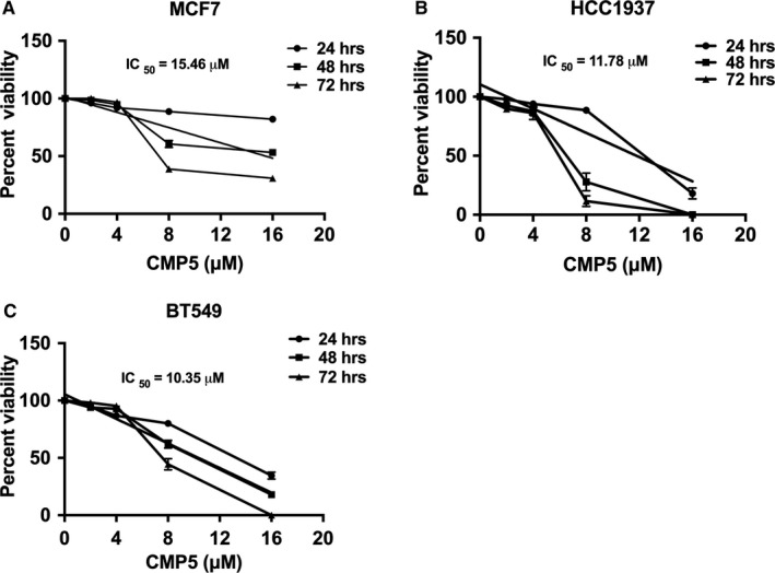
Protein arginine methyltransferase 5 (PRMT5) activity is dysregulated in many aggressive cancers and its enhanced levels are associated with increased tumour growth and survival. However, the role of PRMT5 in breast cancer remains underexplored. In this study, we show that PRMT5 is overexpressed in breast cancer cell lines, and that it promotes WNT/β-CATENIN proliferative signalling through epigenetic silencing of pathway antagonists, DKK1 and DKK3, leading to enhanced expression of c-MYC, CYCLIN D1 and SURVIVIN. Through chromatin immunoprecipitation (ChIP) studies, we found that PRMT5 binds to the promoter region of WNT antagonists, DKK1 and DKK3, and induces symmetric methylation of H3R8 and H4R3 histones. Our findings also show that PRMT5 inhibition using a specific small molecule inhibitor, compound 5 (CMP5), reduces PRMT5 recruitment as well as methylation of H3R8 and H4R3 histones in the promoter regions of DKK1 and DKK3, which consequently results in reduced expression CYCLIN D1 and SURVIVIN. Furthermore, CMP5 treatment either alone or in combination with 5-Azacytidine and Trichostatin A restored expression of DKK1 and DKK3 in TNBCs. PRMT5 inhibition also altered the growth characteristics of breast cancer cells and induced their death. Collectively, these results show that PRMT5 controls breast cancer cell growth through epigenetic silencing of WNT/β-CATENIN pathway antagonists, DKK1 and DKK3, resulting in up-regulation of WNT/β-CATENIN proliferative signalling.

Keywords: PRMT5; WNT/β-CATENIN proliferative signalling; breast cancer; epigenetic silencing; tumour suppressors.


Conflict of interest statement

The authors declare that they have no conflict of interest with the contents of this article.

Figures

FIGURE 1
FIGURE 1
Expression of PRMT5 and WNT/‐CATENIN target genes is elevated and that of WNT/‐CATENIN antagonists, DKK1 and DKK3, is down‐regulated in breast cancer cells. mRNA levels of PRMT5 (A), WNT/‐CATENIN target genes including CYCLIN D1, CYCLIN D3, c‐MYC and SURVIVIN (C) WNT/‐CATENIN antagonists AXIN1, AXIN2, and WIF1 (D), DKK1‐4 (E) and SFRP2‐5 (F) in normal HMECs or MCF7, HCC1937 and BT549 cells were detected by real‐time RT‐PCR using 2 µg of total RNA. The experiment was performed twice with three technical replicates, and ‐ACTIN was used as internal control. The results are represented as mean ± SD. RIPA extracts (20 µg) from either normal HMECs or breast cancer cells were analysed by SDS‐PAGE to detect PRMT5, CYCLIN D1, CYCLIN D3, c‐MYC and SURVIVIN (B) as well as WNT/‐CATENIN antagonists, DKK1 and DKK3 (G) by Western blotting. ‐ACTIN was used as loading control. *** indicates P values < 10−3
FIGURE 2
FIGURE 2
PRMT5 epigenetically suppresses expression of WNT/‐CATENIN antagonists, DKK1 and DKK3 in breast cancer cells. ChIP assays were performed using cross‐linked chromatin from either MCF7 (A), HCC1937 (B) or BT549 (C) cells. Immunoprecipitation of nucleoprotein complexes was performed using pre‐immune (PI), anti‐PRMT5, anti‐H3(Me2)R8 or anti‐H4(Me2)R3 antibodies, and the purified DNA was used to detect the promoter sequences of DKK1 and DKK3 by real‐time PCR using specific primers and probe sets. ChIP assays were repeated two times with three technical replicates. The results are represented as mean ± SD. *** indicates P values < 10−3, ** indicates P values < 10−2 and * indicates P value < 10−1
FIGURE 3
FIGURE 3
PRMT5 inhibition induces transcriptional derepression of WNT/‐CATENIN antagonists DKK1 and DKK3 in TNBC cells. mRNA levels of DKK1 and DKK3 were analysed by real‐time RT‐PCR using 2μg of total RNA extracted from BT549 (A) and HCC1937 (B) treated with increasing amounts of CMP5 (0, 3, 6, 9, 12 μmol/L) for 48 h and 24 h, respectively. (C) Protein levels of DKK1 and DKK3 were analysed by ELISA after collecting the culture media from either DMSO‐ or CMP5‐treated BT549 cells for 48 h. (D) mRNA levels of DKK1 and DKK3 were analysed by real‐time RT‐PCR using 2μg of total RNA extracted from HCC1937 cells treated with 5‐Azacytidine (24 μmol/L) alone or in combination with CMP5 (9 μmol/L) for 24 h. (E) mRNA levels of DKK1 and DKK3 were analysed by real‐time RT‐PCR using 2 μg of total RNA extracted from HCC1937 cells treated with suboptimal concentration of CMP5 (6 μmol/L) in combination with TSA (100 nmol/L) and 5‐Azacytidine (10 μmol/L) for the indicated time intervals. All the experiments were repeated two times with three technical replicates. The results are represented as mean ± SD. *** indicates P values < 10−3 and ** indicates P values < 10−2
FIGURE 4
FIGURE 4
PRMT5 inhibition down‐regulates expression of CYCLIN D1 and SURVIVIN in TNBC cells. HCC1937 (A) and BT549 (B) TNBC cells were treated with increasing amounts of CMP5 (0, 3, 6, 9, 12 μmol/L), and total RNA was extracted 24 h and 48 h post‐treatment, respectively. Levels of WNT/‐CATENIN target genes were analysed by real‐time RT‐PCR using gene‐specific primers and probe sets. The experiment was repeated two times with three technical replicates and ‐ACTIN was used as an internal control. The data shown in the graph represent the mean for each concentration SD. Approximately 20 µg of RIPA extracts from either treated or non‐treated HCC1937 (C) and BT549 (D) cells were analysed by immunoblotting using the indicated antibodies. ⍺‐TUBULIN was detected to show equal loading. *** indicates P values < 10−3, and ** indicates P value < 10−2
FIGURE 5
FIGURE 5
PRMT5 inhibition alters its recruitment and symmetric dimethylation of histones, H3R8 and H4R3 on promoter regions of DKK1 and DKK3. ChIP assay was performed to detect recruitment of PRMT5 and enrichment of its epigenetic marks in the promoter region of WNT/‐CATENIN antagonists as described in Figure 3. Cross‐linked chromatin from either DMSO‐ or CMP5‐treated BT549 (A and B) and HCC1937 (C and D) cells was immunoprecipitated using PI or the indicated immune antibodies. DKK1 and DKK3 promoter sequences of were detected by real‐time PCR using specific primers and probe sets. The experiment was repeated two times with three technical replicates, and data in each graph represent the mean ± SD. *** indicates P values < 10−3, and ** indicates P value < 10−2
FIGURE 6
FIGURE 6
PRMT5 inhibition reduces the viability of breast cancer cells in vitro. MCF7 (A), HCC1937 (B) and BT549 (C) were incubated with different concentrations of CMP5 for 24, 48 and 72 h. Cell viability was measured by Trypan blue dye exclusion assay, and data represented in each graph is from two biological replicates with three technical replicates
FIGURE 7
FIGURE 7
PRMT5 inhibition reduces migration, invasion and induces apoptosis of breast cancer cells. (A) Migration of breast cancer cells was evaluated using Matrigel coated Boyden chamber. Breast cancer cells were treated with either DMSO or CMP5 for 48 h (MCF7 and BT549) or 24 h (HCC1937) as described in Materials and Methods. The migrated cells were stained with crystal violet, and the photomicrographs were captured using an inverted microscope with a magnification of 100×. (B) The percentage of migrated cells was determined using ImageJ software, and the migration capacity was normalized to that of DMSO‐treated cells. (C) Invasion of breast cancer cells was assessed using modified Boyden chamber. MCF7, HCC1937 and BT549 cells were treated with either DMSO or CMP5 for 24 h or 48 h as described in Materials and Methods section. The percentage of invading cells was determined after staining with crystal violet using an inverted microscope with a magnification of 100X. (D) Membrane‐associated invading cells were quantified using ImageJ software, and the per cent invasion was determined in comparison to DMSO‐treated cells. (E) HCC1937 and BT549 cells were treated with different concentrations of CMP5 as indicated, and the number of dead cells was measured by FACS analysis after staining cells with ANNEXIN V and propidium iodide (PI). (F) The average result from the two biological replicates used in E is represented as mean ± SD. All experiments were carried out twice in duplicates, and the data are represented as mean ± SD. *** indicates P values < 10−3

Similar articles

Cited by

References

    1. Siegel RL, Miller KD, Jemal A. Cancer Statistics, 2017. Cancer J Clin. 2017;67(1):7‐30. 10.3322/caac.21387 - DOI - PubMed
    1. World Cancer Report 2014 . World Cancer Report. International Agency for Research on Cancer; 2014.
    1. Temian DC, Pop LA, Irimie AI, Berindan‐Neagoe I. The epigenetics of triple‐negative and basal‐like breast cancer: current knowledge. J Breast Cancer. 2018;21(3):233‐243. 10.4048/jbc.2018.21.e41 - DOI - PMC - PubMed
    1. Pasculli B, Barbano R, Parrella P. Epigenetics of breast cancer: Biology and clinical implication in the era of precision medicine. Semin Cancer Biol. 2018;51:22‐35. 10.1016/j.semcancer.2018.01.007 - DOI - PubMed
    1. Cava C, Bertoli G, Castiglioni I. Integrating genetics and epigenetics in breast cancer: biological insights, experimental, computational methods and therapeutic potential. BMC Syst Biol. 2015;9:62 10.1186/s12918-015-0211-x - DOI - PMC - PubMed

Publication types

MeSH terms