. 2020 Aug 25;19(4):348-361.
doi: 10.18502/ijaai.v19i4.4110.

Fibroblast Growth Factor 2 Augments Transforming Growth Factor Beta 1 Induced Epithelial-mesenchymal Transition in Lung Cell Culture Model

Affiliations

Fibroblast Growth Factor 2 Augments Transforming Growth Factor Beta 1 Induced Epithelial-mesenchymal Transition in Lung Cell Culture Model

Lamis M F El-Baz et al. Iran J Allergy Asthma Immunol. .

Abstract

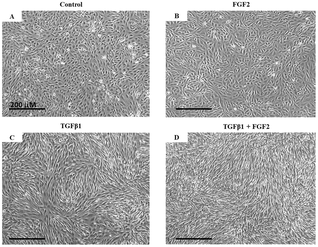
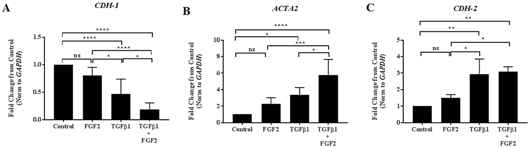
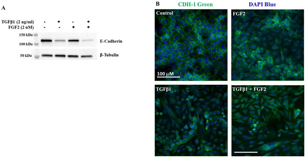
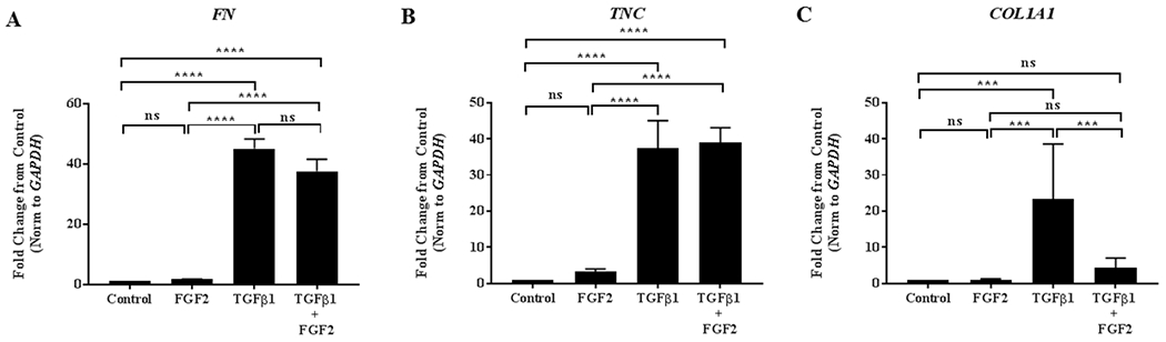
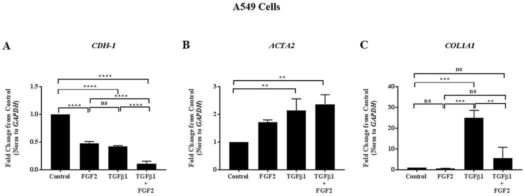
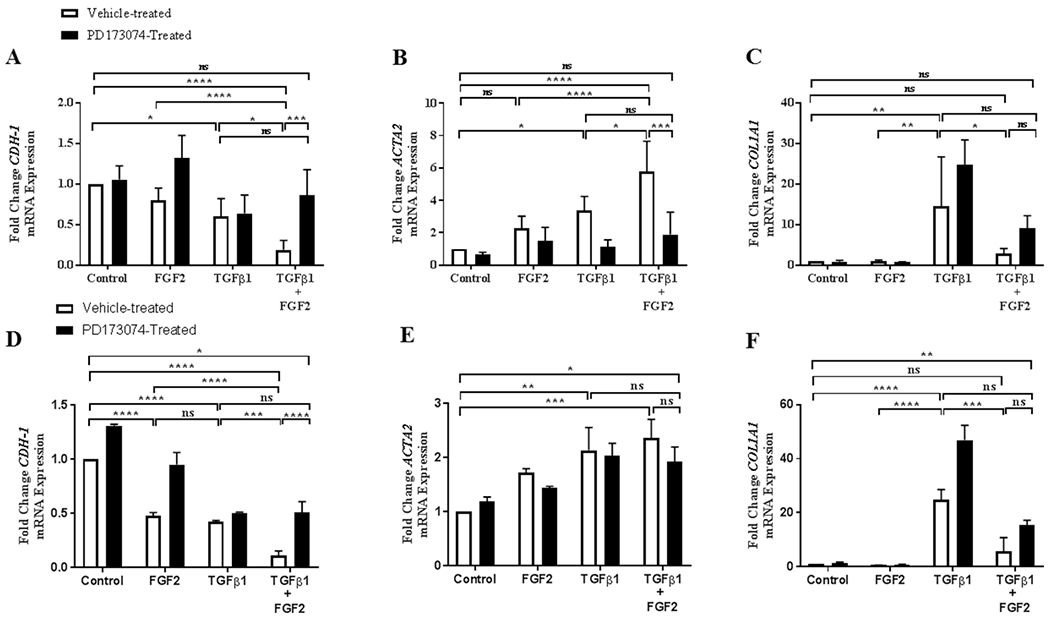
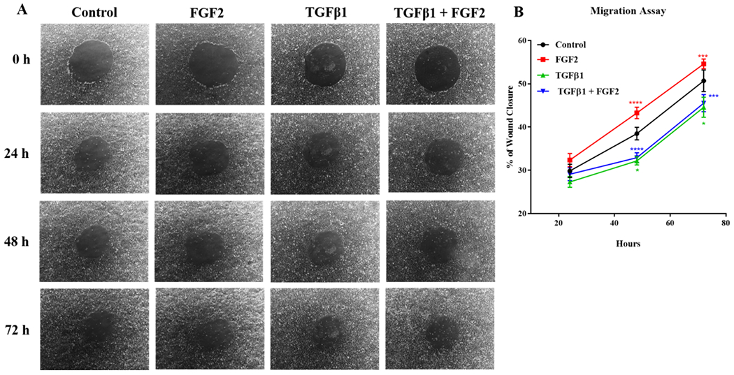
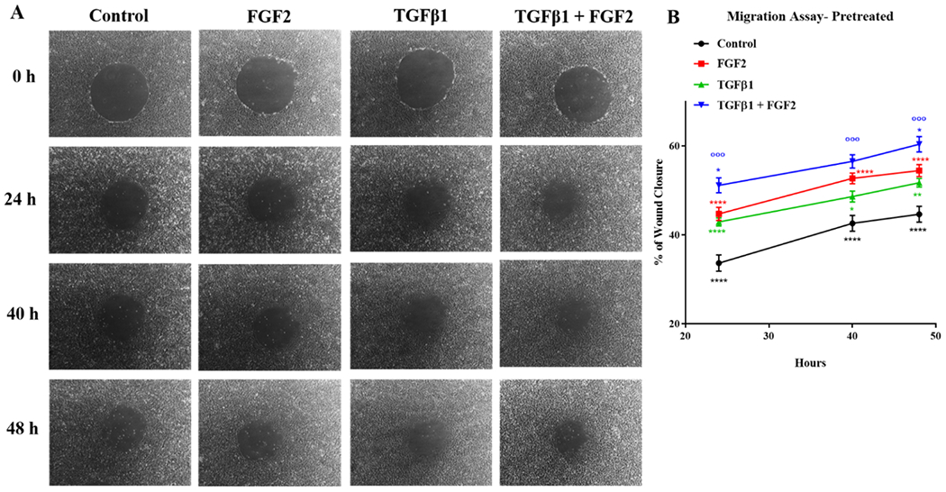
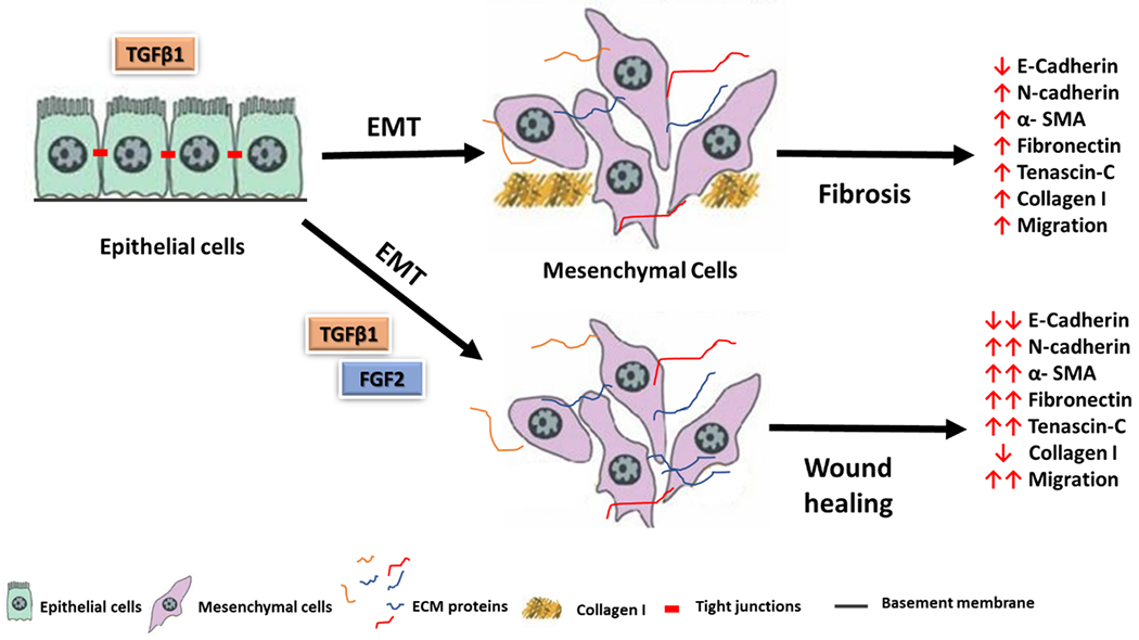
Impaired lung epithelial cell regeneration following injury may contribute to the development of pulmonary fibrosis. Epithelial-mesenchymal transition (EMT) is a critical event in embryonic development, wound healing following injury, and even cancer progression. Previous studies have shown that the combination of transforming growth factor beta-1 (TGFβ1) and fibroblast growth factor 2 (FGF2) induces EMT during cancer metastasis. However, this synergy remains to be elucidated in inducing EMT associated with wound healing after injury. We set out this study to determine the effect of fibroblast growth factor 2 (FGF2) on TGFβ1-induced EMT in the human lung epithelium. BEAS-2B and A549 cells were treated with TGFβ1, FGF2, or both. EMT phenotype was investigated morphologically and by measuring mRNA expression levels; using quantitative real-time PCR. E-cadherin expression was assayed by western blot and immunofluorescence staining. Cell migration was confirmed using a wound-healing assay. TGFβ1 induced a morphological change and a significant increase in cell migration of BEAS-2B cells. TGFβ1 significantly reduced E-cadherin (CDH1) mRNA expression and markedly induced expression of N-cadherin (CDH2), tenascin C (TNC), fibronectin (FN), actin alpha 2 (ACTA2), and collagen I (COL1A1). While FGF2 alone did not significantly alter EMT gene expression, it enhanced TGFβ1-induced suppression of CDH1 and upregulation of ACTA2, but not TNC, FN, and CDH2. FGF2 significantly inhibited TGFβ1-induced COL1A1 expression. Furthermore, FGF2 maintained TGFβ1-induced morphologic changes and increased the migration of TGFβ1-treated cells. This study suggests a synergistic effect between TGFβ1 and FGF2 in inducing EMT in lung epithelial cells, which may play an important role in wound healing and tissue repair after injury.

Keywords: Epithelial cells; Epithelial-mesenchymal transition; Fibroblast growth factor 2; Lung injury; Transforming growth factor beta1.


Conflict of interest statement

Conflict of interest

No conflicts of interest, financial or otherwise, are declared by the authors.

Figures

Figure 1.
Figure 1.. Morphological changes induced by TGFβ1 and FGF2.
BEAS-2B cells were grown in complete growth media (Control) or stimulated with TGFβ1 (2 ng/ml), FGF2 (2 nM) + heparin sulphate (1 nM), or TGFβ1 + FGF2 + heparin sulphate for 4 days. Representative phase-contrast images (10× original magnification) show the morphological change of BEAS-2B cells from cobblestone-like shape as in the control (A) and FGF2 treatment alone (B) to the fibroblast-like shape in the presence of TGFβ1 (C) which is unaltered after adding FGF2 (D).
Figure 2.
Figure 2.. FGF2 enhances TGFβ1-induced EMT gene expression in BEAS-2B cells.
BEAS-2B cells were incubated for 4 days in the absence or presence of 2 ng/ml TGFβ1 alone, FGF2 (2 nM) + heparin sulphate (1 nM), or TGFβ1+ FGF2 + heparin. Quantitative real-time PCR analysis was performed for CDH1 (A), ACTA2 (B) and CDH2 (C). ΔCt values were normalized to GAPDH and expressed as fold change from untreated controls. Statistical significance was determined by one-way ANOVA followed by Tukey’s multiple comparisons test; ns = not significant, * indicates p < 0.05, ** indicates p < 0.01, *** indicates p < 0.001, and **** indicates p < 0.0001.
Figure 3.
Figure 3.. TGFβ1 induced decrease in E-Cadherin protein expression is augmented by FGF2.
(A) Total cell lysates from BEAS-2B cells stimulated for 4 days with or without TGFβ1 (2 ng/ml) alone, FGF2 (2 nM) + heparin sulphate (1 nM), or TGFβ1 + FGF2 + heparin, were immunoblotted for E-cadherin. Blots were reprobed for β-tubulin as a loading control. (B) Densitometry for E-cadherin was normalized to β-tubulin and is expressed as fold change from untreated control. Statistical significance was determined by one-way ANOVA followed by Tukey’s multiple comparisons test; ns = not significant, * indicates p < 0.05, ** indicates p < 0.01, and *** indicates p < 0.001. (C) Representative images showing immunofluorescent staining for E-cadherin in BEAS-2B cells stimulated with or without TGFβ1 alone, FGF2alone or TGFβ1 and FGF2 for 4 days. The confocal images were obtained at 20× original magnification for E-cadherin (green) and DAPI (blue).
Figure 4.
Figure 4.. FGF2 inhibits TGFβ1 induced collagen, but not fibronectin or tenascin-C.
BEAS-2B cells were incubated for 4 days in the absence or presence of 2 ng/ml TGFβ1 alone, FGF2 (2 nM) + heparin sulphate (1 nM), or TGFβ1 + FGF2 + heparin. Quantitative real-time PCR analysis was performed for FN (A), TNC (B) and COL1A1 (C). ΔCt values were normalized to GAPDH and expressed as fold change from untreated controls. Statistical significance was determined by one-way ANOVA followed by Tukey’s multiple comparisons test; ns = not significant, *** indicates p < 0.001, and **** indicates p <0.0001.
Figure 5.
Figure 5.. The effect of FGF2 on TGFβ1-induced EMT gene expression is conserved in A549 cells.
A549 cells were grown in complete Ham’s F12 media (Control) or stimulated with TGFβ1 (2 ng/ml), FGF2 (2 nM) + heparin sulphate (1 nM), or TGFβ1 + FGF2 + heparin sulphate for 4 days. Quantitative real-time PCR analysis was performed for CDH1 (A), ACTA2 (B) and COL1A1 (C) ΔCt values were normalized to GAPDH and expressed as fold change from untreated controls. Statistical significance was determined by one-way ANOVA followed by Tukey’s multiple comparisons test; ns = not significant, ** indicates p < 0.01, *** indicates p < 0.001, and **** indicates p < 0.0001.
Figure 6.
Figure 6.. The effect of FGF2 on TGFβ1 induced EMT gene expression in lung epithelial cells is blocked by the FGFR-specific tyrosine kinase inhibitor PD173074.
BEAS-2B cells were incubated with TGFβ1 (2 ng/ml), FGF2 (2 nM) + heparin sulphate (1 nM), PD173074 (0.1 μM) alone or FGF2 + heparin + TGFβ1 +/− PD173074 for 4 days. Quantitative real-time PCR analysis was performed for CDH1 (A), ACTA2 (B) and COL1A1 (C). A549 cells were incubated with TGFβ1 (2 ng/ml), FGF2 (2 nM) alone, PD173074 (0.1 μM) alone or FGF2 (2 nM) + TGFβ1 +/− PD173074 for 4 days. Quantitative real-time PCR analysis was performed for CDH1 (D), ACTA2 (E) and COL1A1 (F). ΔCt values were normalized to GAPDH and expressed as fold change from untreated controls. Statistical significance was determined by one-way ANOVA followed by Tukey’s multiple comparisons test; ns = not significant, * indicates p < 0.05, ** indicates p < 0.01, *** indicates p < 0.001, and **** indicates p < 0.0001.
Figure 7.
Figure 7.. FGF2, but not TGFβ1, increases BEAS-2B migration of epithelial cells when added immediately after wounding.
BEAS-2B were grown in complete media to confluence, and 1mm diameter circular wounds were generated and the cells were treated immediately with TGFβ1 (2 ng/ml), FGF2 (2 nM) + heparin sulphate (1 nM), or TGFβ1 + FGF2 + heparin. (A) Representative phase-contrast images (10× original magnification) of the same area were taken immediately after wounding (0 h) as well as 24 h, 48 h and 72 h later. (B) Wound area was imaged at 0 h, 24 h, 48 h and 72 h then the % of wound closure was measured using ImageJ software. Statistical significance was determined by one-way ANOVA followed by Tukey’s multiple comparisons test; Red asterisks mark significant differences for FGF2 vs. TGFβ1, blue asterisks mark significant differences for TGFβ1 + FGF2 vs. FGF2, and green asterisks mark significant differences for TGFβ1 vs. control. * indicates p < 0.05, *** indicates p < 0.001 and **** indicates p < 0.0001.
Figure 8.
Figure 8.. Pre-treatment with FGF2 and TGFβ1 significantly increases migration rates of BEAS-2B cells after wounding.
BEAS-2B cells were treated with TGFβ1 (2 ng/ml), FGF2 (2 nM) + heparin sulphate (1 nM), or TGFβ1 + FGF2 + heparin for 3 days. The cells then wounded and the % of wound closure was measured at 0 h, 24 h, 40 h and 48 h later. (A) Representative phase contrast images (10× original magnification) of the same area were taken immediately after wounding (0 h) as well as 24 h, 40 h and 48 h later. (B) Statistical significance was determined by one-way ANOVA followed by Tukey’s multiple comparisons test; Red asterisks mark significant differences for FGF2 vs. control, blue asterisks mark significant differences for TGFβ1 + FGF2 vs. FGF2, blue circles mark significant differences for TGFβ1 + FGF2 vs. TGFβ1, green asterisks mark significant differences for TGFβ1 vs. control and black asterisks mark significant differences for control vs TGFβ1 + FGF2. * indicates p < 0.05, ** indicates p < 0.01, *** indicates p < 0.001, and **** indicates p < 0.0001.
Figure 9.
Figure 9.
A schematic diagram of FGF2 and TGFβ1 for the induction of epithelial-mesenchymal transition in lung epithelial cells. TGFβ1: transforming growth factor beta 1, FGF2: fibroblast growth factor 2, EMT; epithelial-mesenchymal transition, α-SMA; alpha smooth muscle actin, ECM; extracellular matrix proteins.

Similar articles

Cited by

References

    1. Desai O, Winkler J, Minasyan M, Herzog EL. The Role of Immune and Inflammatory Cells in Idiopathic Pulmonary Fibrosis. Front Med [Internet]. 2018March20 [cited 2019 Aug 27];5:43. Available from: http://www.ncbi.nlm.nih.gov/pubmed/29616220 - PMC - PubMed
    1. King TE, Pardo A, Selman M. Idiopathic pulmonary fibrosis. Lancet [Internet]. 2011December3 [cited 2019 Aug 4];378(9807):1949–61. Available from: http://www.ncbi.nlm.nih.gov/pubmed/21719092 - PubMed
    1. Yang J, Wheeler SE, Velikoff M, Kleaveland KR, Lafemina MJ, Frank JA, et al.Activated alveolar epithelial cells initiate fibrosis through secretion of mesenchymal proteins. Am J Pathol. 2013November;183(5):1559–70. - PMC - PubMed
    1. Li M, Luan F, Zhao Y, Hao H, Zhou Y, Han W, et al.Epithelial-mesenchymal transition: An emerging target in tissue fibrosis. Exp Biol Med (Maywood) [Internet]. 2016January [cited 2018 Mar 9];241(1):1–13. Available from: http://www.ncbi.nlm.nih.gov/pubmed/26361988 - PMC - PubMed
    1. Rout-Pitt N, Farrow N, Parsons D, Donnelley M. Epithelial mesenchymal transition (EMT): A universal process in lung diseases with implications for cystic fibrosis pathophysiology [Internet]. Vol. 19, Respiratory Research. 2018. [cited 2019 Sep 24]. p. 136. Available from: 10.1186/s12931-018-0834-8 - DOI - PMC - PubMed

MeSH terms