. 2021 Mar 23;143(12):1242-1255.
doi: 10.1161/CIRCULATIONAHA.120.051889. Epub 2021 Jan 19.

Isolevuglandin-Modified Cardiac Proteins Drive CD4+ T-Cell Activation in the Heart and Promote Cardiac Dysfunction

Affiliations

Isolevuglandin-Modified Cardiac Proteins Drive CD4+ T-Cell Activation in the Heart and Promote Cardiac Dysfunction

Njabulo Ngwenyama et al. Circulation. .

Erratum in

Abstract

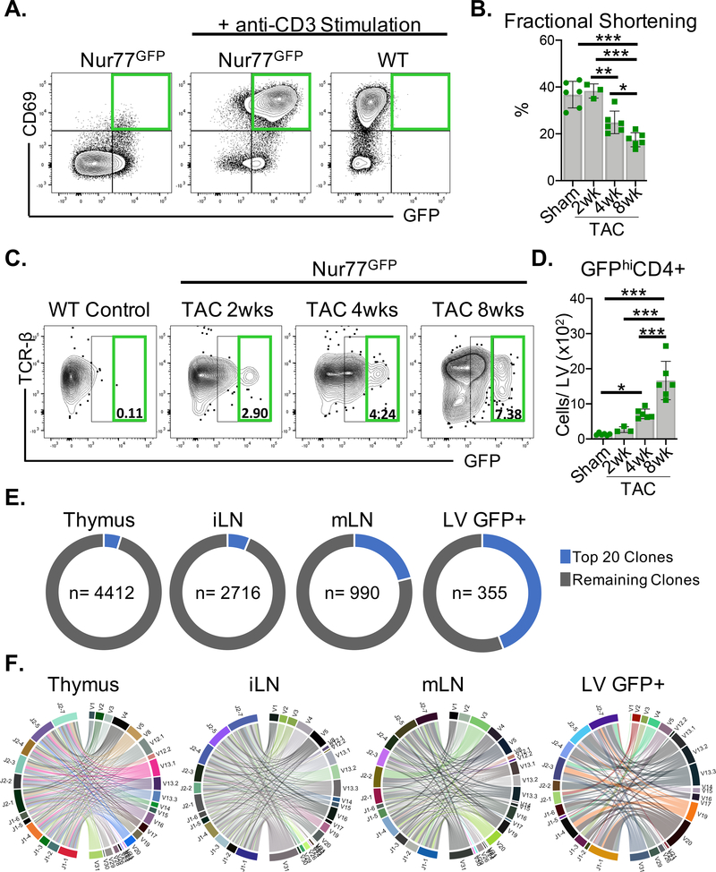
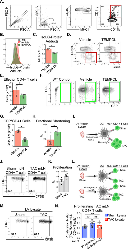
Background: Despite the well-established association between T-cell-mediated inflammation and nonischemic heart failure, the specific mechanisms triggering T-cell activation during the progression of heart failure and the antigens involved are poorly understood. We hypothesized that myocardial oxidative stress induces the formation of isolevuglandin (IsoLG)-modified proteins that function as cardiac neoantigens to elicit CD4+ T-cell receptor (TCR) activation and promote heart failure.

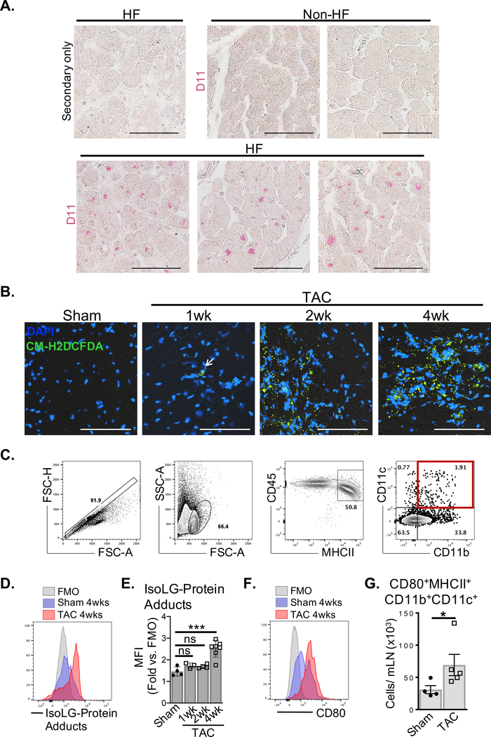
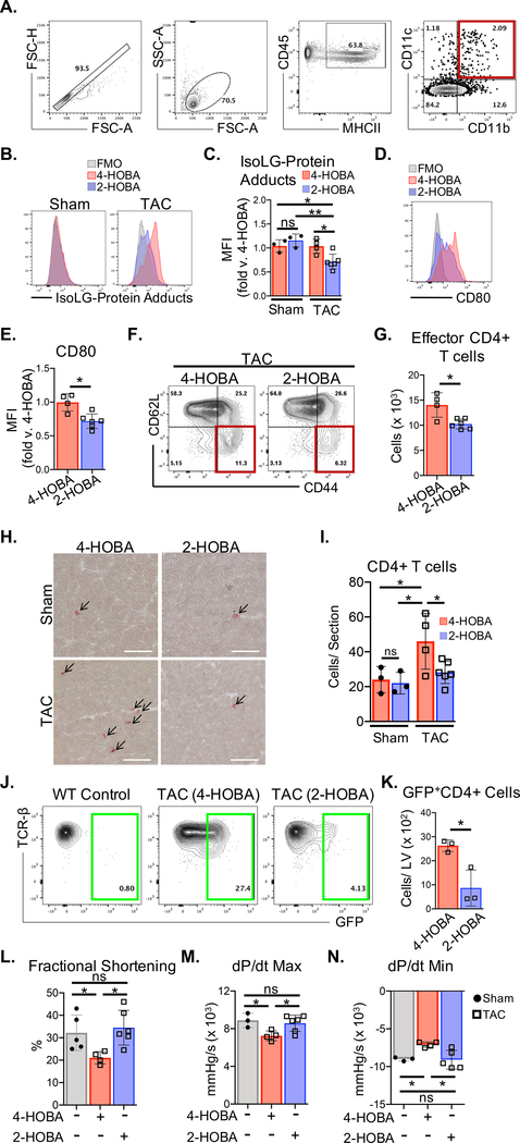
Methods: We used transverse aortic constriction in mice to trigger myocardial oxidative stress and T-cell infiltration. We profiled the TCR repertoire by mRNA sequencing of intramyocardial activated CD4+ T cells in Nur77GFP reporter mice, which transiently express GFP on TCR engagement. We assessed the role of antigen presentation and TCR specificity in the development of cardiac dysfunction using antigen presentation-deficient MhcII-/- mice and TCR transgenic OTII mice that lack specificity for endogenous antigens. We detected IsoLG protein adducts in failing human hearts. We also evaluated the role of reactive oxygen species and IsoLGs in eliciting T-cell immune responses in vivo by treating mice with the antioxidant TEMPOL and the IsoLG scavenger 2-hydroxybenzylamine during transverse aortic constriction, and ex vivo in mechanistic studies of CD4+ T-cell proliferation in response to IsoLG-modified cardiac proteins.

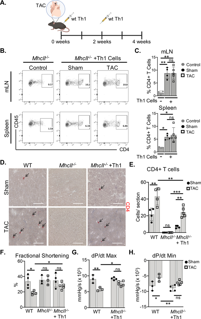
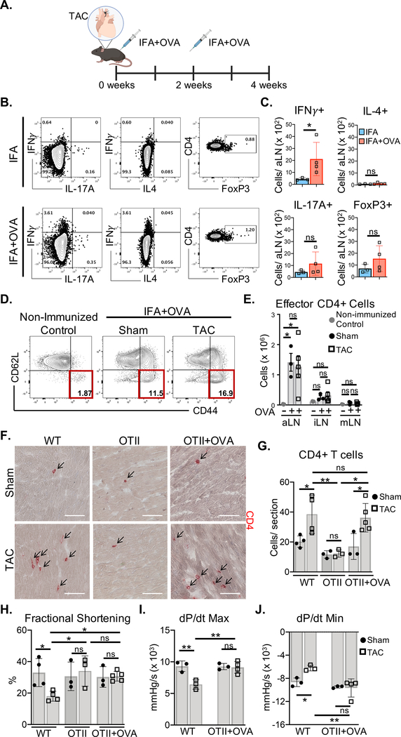
Results: We discovered that TCR antigen recognition increases in the left ventricle as cardiac dysfunction progresses and identified a limited repertoire of activated CD4+ T-cell clonotypes in the left ventricle. Antigen presentation of endogenous antigens was required to develop cardiac dysfunction because MhcII-/- mice reconstituted with CD4+ T cells and OTII mice immunized with their cognate antigen were protected from transverse aortic constriction-induced cardiac dysfunction despite the presence of left ventricle-infiltrated CD4+ T cells. Scavenging IsoLGs with 2-hydroxybenzylamine reduced TCR activation and prevented cardiac dysfunction. Mechanistically, cardiac pressure overload resulted in reactive oxygen species-dependent dendritic cell accumulation of IsoLG protein adducts, which induced robust CD4+ T-cell proliferation.

Conclusions: Our study demonstrates an important role of reactive oxygen species-induced formation of IsoLG-modified cardiac neoantigens that lead to TCR-dependent CD4+ T-cell activation within the heart.

Keywords: heart failure; inflammation; isolevuglandin; oxidative stress.


Conflict of interest statement

Conflict of Interest Disclosures

None

Figures

Figure 1.
Figure 1.. CD4+ T cells are activated via the TCR in the LV in response to TAC.
(A) CD4+ T cells isolated from wt or Nur77GFP mice were stimulated with immobilized anti-CD3 (5μg/mL) in vitro for 72 hours where indicated. GFP expression and the control T cell activation marker CD69 were evaluated by FACS. (B-F) Nur77GFP mice underwent Sham surgery for 8wks and TAC surgery for 2–8wks. (B) Transthoracic echocardiography was used to measure LV fractional shortening. (C-D) Leukocytes were isolated from the LV and the total number of GFPhiCD4+ T cells was quantified by FACS. (E-F) Bulk TCR clonal analysis after 8wks of TAC surgery (n=3 TAC Nur77GFP mice pooled) was performed on 5000 CD4+ T cells sorted from the thymus, inguinal lymph nodes (iLN), mediastinal lymph nodes (mLN) and 5000 GFP+CD4+ T cells sorted from the LV by next generation RNA sequencing. (E) The frequency of the most abundant 20 clones from each site are represented in the pie graph along with total number (n) of unique clones identified. (F) CD4+ T cell clonotypes are represented in chord diagrams, with ribbons proportionately scaled by V-J pair frequency connecting segment pairs. Error bars represent mean ± SD. (* p<0.05, ** p<0.01, *** p<0.001; one-way ANOVA test).
Figure 2.
Figure 2.. MhcII−/− mice reconstituted with Th1 cells do not develop cardiac dysfunction in response to TAC.
(A) Wt Th1 cells were differentiated in vitro and transferred to MhcII−/− mice via intraperitoneal injection, 2 days and 2 weeks after Sham and TAC. surgery. (B-C) The reconstitution with CD4+ T cells was evaluated in the mediastinal lymph nodes (mLNs) and spleen by FACS, and compared to control MhcII−/− mice that did not receive Th1 cells. (D-E) Frozen LV tissue sections isolated from wt, MhcII−/−, and MhcII−/− mice reconstituted with Th1 cells, 4 weeks after Sham or TAC surgery were used to determine CD4+ T cell LV infiltration. (F) Transthoracic echocardiography was used to measure LV fractional shortening from wt, MhcII−/−, and MhcII−/− mice reconstituted with Th1 cells. (G-H) LV hemodynamic measurements were acquired to determine dP/dt max and dP/dt min as parameters of cardiac contractility and relaxation respectively in MhcII−/− mice reconstituted with Th1 cells compared to untreated wt mice. Scale bars: 100μm. Error bars represent mean ± SD. (* p<0.05, ** p<0.01, *** p<0.001; one-way ANOVA test, two-way ANOVA with two categorical grouping variables).
Figure 3.
Figure 3.. Activated OVA-specific CD4+ T cells recruited to the LV of OTII mice do not mediate cardiac dysfunction in response to TAC.
(A) OTII mice were immunized with ovalbumin peptide (OVA) emulsified in Incomplete Freund’s adjuvant (IFA), 2 days and 2 weeks after Sham and TAC surgery. (B-C) Cytokine production and FoxP3 expression was evaluated in CD4+ T cells from the draining axillary lymph nodes (aLNs) of immunized OTII mice by intracellular staining after 3–5hrs of PMA/ionomycin stimulation. (D-E) 4 weeks after surgery CD4+ T cell activation was evaluated by quantifying the number of effector (CD62Llo CD44hi) CD4+ T cells in the draining axillary lymph nodes (aLN) as well as the inguinal lymph nodes (iLN) and mediastinal lymph nodes (mLNs) by FACS. (F-G) IHC in frozen LV tissue sections was used to determine CD4+ T cell infiltration in wt mice, OTII mice, and OTII mice immunized with OVA, 4 weeks after Sham and TAC surgeries. (H) Transthoracic echocardiography was used to measure LV fractional shortening (I-J) LV hemodynamic measurements were acquired to determine dP/dt max and dP/dt min in OTII mice immunized with OVA and wt mice, 4 weeks after Sham and TAC surgeries. Scale bars: 100 μm. Error bars represent mean ± SD. (* p<0.05, ** p<0.01; one-way ANOVA test, two-way ANOVA with two categorical grouping variables).
Figure 4.
Figure 4.. HF is associated with increased accumulation of Isolevuglandin (IsoLG)-protein adducts.
(A) Fixed LV tissue sections from non-HF (n=2) and end-stage nonischemic HF patients (n=3) were probed for the presence of IsoLG-protein adducts with D11 scfv recombinant antibody. (B) Frozen LV tissue sections from wt Sham (4wks post-surgery) and TAC (1wk, 2wk and 4wk post-surgery) mice were probed for intracellular ROS by CM-H2DCFDA. (Images representative of n≥3 mice each) (C) CD45+MHCII+CD11b+CD11c+ DCs isolated from the mLNs of wt Sham and TAC mice were identified by FACS. (D-E) DCs mice were then probed for intracellular IsoLG-protein adducts with D11 scfv recombinant antibody and quantified. (F-G) The total number of mature DCs expressing CD80 in wt Sham and TAC mLNs was quantified by FACS. Scale bars: 100μm. Error bars represent mean ± SD. (* p<0.05, *** p<0.001; Mann-Whitney test, one-way ANOVA test for comparison of 3 or more groups).
Figure 5.
Figure 5.. Oxidative stress during TAC promotes LV CD4+ T cell activation in response to IsoLG-modified proteins.
(A-H) Nur77GFP mice were treated orally with the antioxidant 4-hydroxy-TEMPO (TEMPOL) administered ad libitum in the drinking water (1mM) for 4 weeks post TAC surgery. (A-C) CD45+MHCII+CD11b+CD11c+ DCs were isolated from the heart-draining mLNs of vehicle and TEMPOL treated mice and probed for intracellular IsoLG-protein adducts by FACS using D11 scfv recombinant antibody. (D-E) CD62LloCD44hi effector CD4+ T cells in the mLNs were quantified by FACS. (F-G) Total GFP+CD4+ T cells from the whole LV of vehicle and TEMPOL treated Nur77GFP mice were quantified by FACS (H) Transthoracic echocardiography was used to measure LV fractional shortening. (I) CD4+ T cells were purified from the mLN of wt Sham and TAC surgery mice, and cocultured with BMDCs pulsed with 100μg/mL of Sham LV protein lysate pretreated with IsoLGs (100μM). (J-K) CD4+ T cell proliferation was quantified after 72hrs based on the CFSE dilution in mLN CD4+ T cells. (L-N) BMDCs were pulsed with 100μg/mL of Sham or TAC LV protein lysate, or Sham LV lysate pretreated with 100μM of IsoLG and co-cultured with CFSE-labeled mLN CD4+ T cells from Sham and TAC surgery mice. Normalized data are represented as the fold change in proliferation of TAC mLN CD4+ T cells relative to Sham mLN CD4+ T cells in response to the indicated LV protein lysate conditions. Error bars represent mean ± SD. (* p<0.05, ** p<0.01; Mann-Whitney test, One-way ANOVA test for comparison of 3 or more groups).
Figure 6.
Figure 6.. The IsoLG scavenger 2-HOBA reduces CD4+ T cell activation and ameliorates cardiac dysfunction due to TAC.
Mice were treated orally with the IsoLG scavenger 2-HOBA and the control drug 4-HOBA administered ad libitum in the drinking water (1g/L) for 4 weeks after TAC surgery. (A) CD45+MHCII+CD11b+CD11c+ DCs were isolated from the mLNs of wt mice and (B-C) intracellular IsoLG-protein adduct accumulation was measured using D11 scfv recombinant antibody, and (D-E) CD80 surface expression was quantified by FACS. (F-G) Total CD62LloCD44hi effector CD4+ T cells in the mLNs of wt TAC mice were quantified by FACS. (H-I) IHC was used to determine CD4+ T cell infiltration in frozen LV tissue sections from wt mice. (J-K) Total GFP+CD4+ T cells from LV tissues of Nur77GFP mice treated with 2-HOBA and 4-HOBA were quantified by FACS, 4 weeks after TAC surgery. (L) Transthoracic echocardiography was used to measure LV fractional shortening and (M-N) LV hemodynamic measurements were acquired to determine dP/dt max and dP/dt min, comparing untreated wt Sham mice to wt TAC mice treated with 4-HOBA and 2-HOBA. Scale bars: 100μm. Error bars represent mean ± SEM. (* p<0.05, ** p<0.01; Mann-Whitney test, One-way ANOVA test for comparison of 3 or more groups).

Similar articles

Cited by

References

    1. Carrillo-Salinas FJ, Ngwenyama N, Anastasiou M, Kaur K and Alcaide P. Heart Inflammation: Immune Cell Roles and Roads to the Heart. Am J Pathol. 2019;189:1482–1494. - PMC - PubMed
    1. Nevers T, Salvador AM, Grodecki-Pena A, Knapp A, Velazquez F, Aronovitz M, Kapur NK, Karas RH, Blanton RM and Alcaide P. Left Ventricular T-Cell Recruitment Contributes to the Pathogenesis of Heart Failure. Circ Heart Fail. 2015;8:776–87. - PMC - PubMed
    1. Nevers T, Salvador AM, Velazquez F, Ngwenyama N, Carrillo-Salinas FJ, Aronovitz M, Blanton RM and Alcaide P. Th1 effector T cells selectively orchestrate cardiac fibrosis in nonischemic heart failure. The Journal of Experimental Medicine. 2017;214:3311–3329. - PMC - PubMed
    1. Salvador AM, Nevers T, Velazquez F, Aronovitz M, Wang B, Abadia Molina A, Jaffe IZ, Karas RH, Blanton RM and Alcaide P. Intercellular Adhesion Molecule 1 Regulates Left Ventricular Leukocyte Infiltration, Cardiac Remodeling, and Function in Pressure Overload-Induced Heart Failure. J Am Heart Assoc. 2016;5:e003126. - PMC - PubMed
    1. Laroumanie F, Douin-Echinard V, Pozzo J, Lairez O, Tortosa F, Vinel C, Delage C, Calise D, Dutaur M, Parini A and Pizzinat N. CD4+ T cells promote the transition from hypertrophy to heart failure during chronic pressure overload. Circulation. 2014;129:2111–24. - PubMed

Publication types