

Endoscopic visualization-assisted corneal bee sting removal
- PMID: 33463602
- PMCID: PMC7933875
- DOI: 10.4103/ijo.IJO_1161_20
Endoscopic visualization-assisted corneal bee sting removal
Abstract
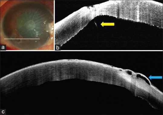
Purpose: Deeply embedded corneal foreign bodies and intrastromal foreign body removal can often be a challenge. The aim of this report was to describe the utility of endoscopy in visualization and removal of an embedded corneal bee stinger.
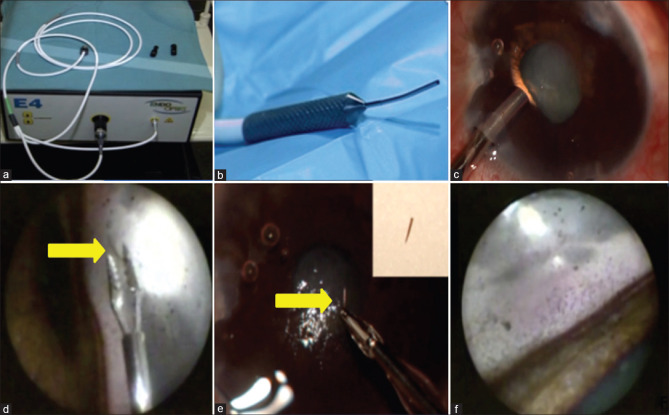
Methods: A 44-year-old male patient developed toxic keratopathy after injury from a bee stinger. On examination, the bee stinger was noted to be deeply embedded in the corneal stroma. A superficial keratectomy was initially attempted; however, the stinger was noted to be intrastromal and protruding into the anterior chamber and could not be removed. An Endoscopy-assisted visualization was used to remove the stinger.
Results: The bee stinger was successfully removed and the patient's vision improved to 20/100 from an initial CFCF (counting fingers close to face) at time of presentation. At the end of 3 months follow-up, there was residual corneal edema along with cataractous changes in the lens as a sequelae of the initial bee sting injury. The patient subsequently underwent an endothelial keratoplasty along with phacoemulsification with intraocular lens implantation and the final BCVA improved to 20/40.
Conclusion: Endoscopyassisted visualisation of anterior chamber and angle structures can be valuable in removal of retained and deeply embedded corneal or intracameral foreign bodies.
Keywords: Bee sting injury; embedded corneal foreign body; endoscopy; toxic keratitis.
Conflict of interest statement
None
Figures
Comment in
-
Commentary: Corneal bee sting injury.Indian J Ophthalmol. 2021 Feb;69(2):426-427. doi: 10.4103/ijo.IJO_2484_20. Indian J Ophthalmol. 2021. PMID: 33463603 Free PMC article. No abstract available.
References
-
- Lin PH, Wang NK, Hwang YS, Ma DH, Yeh LK. Bee sting of the cornea and conjunctiva: Management and outcomes. Cornea. 2011;30:392–4. - PubMed
-
- Arcieri ES, França ET, de Oliveria HB, De Abreu Ferreira L, Ferreira MA, Rocha FJ. Ocular lesions arising after stings by hymenopteran insects. Cornea. 2002;21:328–30. - PubMed
-
- Kim JM, Kang SJ, Kim MK, Wee WR, Lee JH. Corneal wasp sting accompanied by optic neuropathy and retinopathy. Jpn J Ophthalmol. 2011;55:165–7. - PubMed
Publication types
MeSH terms
LinkOut - more resources
Full Text Sources
Other Literature Sources
Medical
Research Materials
