. 2020 Oct;35(10):2070-2081.
doi: 10.1002/jbmr.4110. Epub 2020 Jul 12.

Restriction of Dietary Phosphate Ameliorates Skeletal Abnormalities in a Mouse Model for Craniometaphyseal Dysplasia

Affiliations

Restriction of Dietary Phosphate Ameliorates Skeletal Abnormalities in a Mouse Model for Craniometaphyseal Dysplasia

Yasuyuki Fujii et al. J Bone Miner Res. 2020 Oct.

Abstract

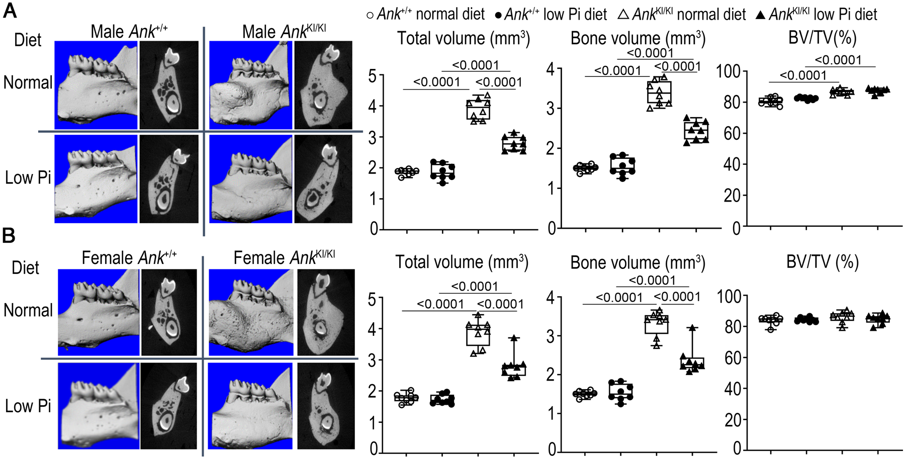
Craniometaphyseal dysplasia (CMD), a rare genetic bone disorder, is characterized by lifelong progressive thickening of craniofacial bones and metaphyseal flaring of long bones. The autosomal dominant form of CMD is caused by mutations in the progressive ankylosis gene ANKH (mouse ortholog Ank), encoding a pyrophosphate (PPi) transporter. We previously reported reduced formation and function of osteoblasts and osteoclasts in a knockin (KI) mouse model for CMD (AnkKI/KI) and in CMD patients. We also showed rapid protein degradation of mutant ANK/ANKH. Mutant ANK protein displays reduced PPi transport, which may alter the inorganic phosphate (Pi) and PPi ratio, an important regulatory mechanism for bone mineralization. Here we investigate whether reducing dietary Pi intake can ameliorate the CMD-like skeletal phenotype by comparing male and female Ank+/+ and AnkKI/KI mice exposed to a low (0.3%) and normal (0.7%) Pi diet for 13 weeks from birth. Serum Pi and calcium (Ca) levels were not significantly changed by diet, whereas PTH and 25-hydroxy vitamin D (25-OHD) were decreased by low Pi diet but only in male Ank+/+ mice. Importantly, the 0.3% Pi diet significantly ameliorated mandibular hyperostosis in both sexes of AnkKI/KI mice. A tendency of decreased femoral trabeculation was observed in male and female Ank+/+ mice as well as in male AnkKI/KI mice fed with the 0.3% Pi diet. In contrast, in female AnkKI/KI mice the 0.3% Pi diet resulted in increased metaphyseal trabeculation. This was also the only group that showed increased bone formation rate. Low Pi diet led to increased osteoclast numbers and increased bone resorption in all mice. We conclude that lowering but not depleting dietary Pi delays the development of craniofacial hyperostosis in CMD mice without severely compromising serum levels of Pi, Ca, PTH, and 25-OHD. These findings may have implications for better clinical care of patients with CMD. © 2020 American Society for Bone and Mineral Research.

Keywords: BONE HISTOMORPHOMETRY; BONE QCT/μCT; GENETIC ANIMAL MODELS; OSTEOBLASTS; OSTEOCLASTS.


Conflict of interest statement

Conflict of Interest:

All authors have no conflict of interest.

Figures

FIG. 1.
FIG. 1.
Restriction of dietary Pi ameliorates hyperostotic mandibles in AnkKI/KI mice. μCT images of 3D reconstruction of mandibles and sagittal plane through furcation of first mandibular molar in A) male Ank+/+ and AnkKI/KI mice; B) female Ank+/+ and AnkKI/KI mice. Histograms showing total volume (TV), bone volume (BV) and bone volume fraction (BV/TV). n=8 per group. p<0.05 indicates statistical significance by one-way ANOVA with Tukey’s test. p-values are indicated above the horizontal bars up to a value of 0.1.
FIG. 2.
FIG. 2.
Effects of dietary Pi on femoral trabecular and cortical bones in Ank+/+ and AnkKI/KI mice. Representative μCT images of internal view of Ank+/+ and AnkKI/KI femurs, 3D reconstruction of trabeculation in metaphysis and cross-sectional slice of cortical bone in diaphysis in A) male Ank+/+ and AnkKI/KI mice; B) female Ank+/+ and AnkKI/KI mice. Histograms show quantitative μCT data of C) male Ank+/+ and AnkKI/KI mice; D) female Ank+/+ and AnkKI/KI mice. n=8 per group. p<0.05 indicates statistical significance by one-way ANOVA with Tukey’s test. p-values are indicated above the horizontal bars up to a value of 0.1.
FIG. 3.
FIG. 3.
Dynamic histomorphometry of Ank+/+ and AnkKI/KI mice exposed to normal and low Pi diet. Representative images of femurs with calcein (green) and alizarin complexone (red) double labeling in A) male Ank+/+ and AnkKI/KI mice; B) female Ank+/+ and AnkKI/KI mice. Quantitative measurement of histomorphometry of C) male Ank+/+ and AnkKI/KI mice; D) female Ank+/+ and AnkKI/KI mice. MS_BS: mineralizing surface per bone surface; MAR: mineral apposition rate; BFR: bone formation rate; AP_BS: alkaline phosphatase-positive labeled surface per bone surface; bone forming surface (AP_L/BS): proportion of AP signal on labeled bone surface; bone lining surface (AP_NL/BS): proportion of AP signal on non-labeled bone surface. p<0.05 indicates statistical significance by one-way ANOVA with Tukey’s test. p- values are indicated above or below the horizontal bars up to a value of 0.1.
FIG. 4.
FIG. 4.
Effects of dietary Pi on bone resorbing activity/surface in Ank+/+ and AnkKI/KI mice. Representative images of femurs with TRAP staining to indicate osteoclast-like cells in A) male Ank+/+ and AnkKI/KI mice; B) female Ank+/+ and AnkKI/KI mice. Quantitative histomorphometry measurements of C) male Ank+/+ and AnkKI/KI mice; D) female Ank+/+ and AnkKI/KI mice. TRAP_BS: fraction of bone surface with TRAP label; TRAP_L/BS: proportion of mineralizing surface that is TRAP-labeled; TRAP_NL/BS: proportion of non-mineralizing surface that is TRAP-labeled. p<0.05 indicates statistical significance by one-way ANOVA with Tukey’s test. p-values are indicated above the horizontal bars up to a value of 0.1. Enlarged images of white square boxes in panels A) and B) showing TRAP labeled surfaces of cortical bones in E) male Ank+/+ and AnkKI/KI mice; F) female Ank+/+ and AnkKI/KI mice. “x” indicates periosteal surface; “*” indicates endosteal surface.
FIG. 5.
FIG. 5.
Effects of reduced Pi concentration on BMSC and BMM cultures derived from Ank+/+ and AnkKI/KI mice. A) AP staining of Ank+/+ and AnkKI/KI BMSCs cultured in osteoblast differentiating medium for 0, 7, 14 and 21 days. AnkKI/KI cultures show reduced AP staining. BMSCs cultured in 0.25 mM Pi showed more AP staining than those cultured in 1 mM Pi medium; B) representative fold-changes of Alpl, Phex, Dmp1 and Spp1 expression in Ank+/+ and AnkKI/KI BMSCs cultured in differentiation medium for 21 days and normalized to expression levels of Ank+/+ BMSCs at day 0, normalized to 18S mRNA from one experiment (n=3). C) Ank+/+ and AnkKI/KI BMMs stained for TRAP in osteoclast differentiating medium for 5 days. Histogram shows numbers of TRAP-stained multinucleated (nuclei ≥ 3) per well; D) measurement of osteoclast surface in pixel diagram. More than 300 osteoclasts per group were measured. Pooled data from three independent experiments. p<0.05 indicates statistical significance by two-way ANOVA with Tukey’s test. p-values are indicated above or below the horizontal bars up to a value of 0.1.

Similar articles

Cited by

References

    1. Elcioglu N, Hall CM. Temporal aspects in craniometaphyseal dysplasia: autosomal recessive type. American journal of medical genetics. Mar 19 1998;76(3):245–51. - PubMed
    1. Ramseyer LT, Leonard JC, Stacy TM. - Bone scan findings in craniometaphyseal dysplasia. - Clinical Nuclear Medicine. 1993;18(2):137–9. - PubMed
    1. Nurnberg P, Thiele H, Chandler D, Hohne W, Cunningham ML, Ritter H, et al. Heterozygous mutations in ANKH, the human ortholog of the mouse progressive ankylosis gene, result in craniometaphyseal dysplasia. Nature genetics. May 2001;28(1):37–41. - PubMed
    1. Reichenberger E, Tiziani V, Watanabe S, Park L, Ueki Y, Santanna C, et al. Autosomal dominant craniometaphyseal dysplasia is caused by mutations in the transmembrane protein ANK. American journal of human genetics. Jun 2001;68(6):1321–6. - PMC - PubMed
    1. Kanaujiya J, Bastow E, Luxmi R, Hao Z, Zattas D, Hochstrasser M, et al. Rapid degradation of progressive ankylosis protein (ANKH) in craniometaphyseal dysplasia. Sci Rep. Oct 24 2018;8(1):15710. Epub 2018/10/26. - PMC - PubMed

Publication types

Supplementary concepts