

Cell-Based High-Throughput Screening Protocol for Discovering Antiviral Inhibitors Against SARS-COV-2 Main Protease (3CLpro)
- PMID: 33464543
- PMCID: PMC7814170
- DOI: 10.1007/s12033-021-00299-7
Cell-Based High-Throughput Screening Protocol for Discovering Antiviral Inhibitors Against SARS-COV-2 Main Protease (3CLpro)
Abstract
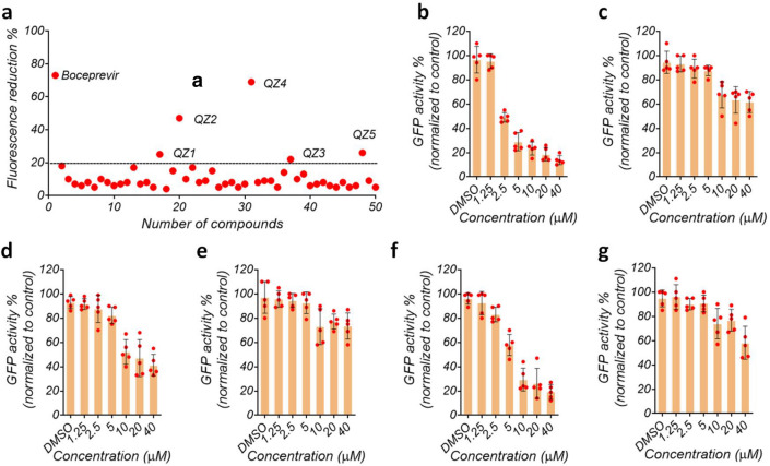
The global public health has been compromised since the severe acute respiratory syndrome coronavirus-2 (SARS-CoV-2) emerged in late December 2019. There are no specific antiviral drugs available to combat SARS-CoV-2 infection. Besides the rapid dissemination of SARS-CoV-2, several variants have been identified with a potential epidemiologic and pathogenic variation. This fact has forced antiviral drug development strategies to stay innovative, including new drug discovery protocols, combining drugs, and establishing new drug classes. Thus, developing novel screening methods and direct-targeting viral enzymes could be an attractive strategy to combat SARS-CoV-2 infection. In this study, we designed, optimized, and validated a cell-based assay protocol for high-throughput screening (HTS) antiviral drug inhibitors against main viral protease (3CLpro). We applied the split-GFP complementation to develop GFP-split-3CLpro HTS system. The system consists of GFP-based reporters that become fluorescent upon cleavage by SARS-CoV-2 protease 3CLpro. We generated a stable GFP-split-3CLpro HTS system valid to screen large drug libraries for inhibitors to SARS-CoV-2 main protease in the bio-safety level 2 laboratory, providing real-time antiviral activity of the tested compounds. Using this assay, we identified a new class of viral protease inhibitors derived from quinazoline compounds that worth further in vitro and in vivo validation.
Keywords: Drug libraries; GFP complementation; High-throughput screening; Protease; SARS-CoV-2.
Conflict of interest statement
The authors declare that they have no conflict of interest with the contents of this article.
Figures


Similar articles
-
Development of a Fluorescence-Based, High-Throughput SARS-CoV-2 3CLpro Reporter Assay.J Virol. 2020 Oct 27;94(22):e01265-20. doi: 10.1128/JVI.01265-20. Print 2020 Oct 27. J Virol. 2020. PMID: 32843534 Free PMC article.
-
3-chymotrypsin-like protease in SARS-CoV-2.Biosci Rep. 2024 Aug 28;44(8):BSR20231395. doi: 10.1042/BSR20231395. Biosci Rep. 2024. PMID: 39036877 Free PMC article. Review.
-
Bisindolylmaleimide IX: A novel anti-SARS-CoV2 agent targeting viral main protease 3CLpro demonstrated by virtual screening pipeline and in-vitro validation assays.Methods. 2021 Nov;195:57-71. doi: 10.1016/j.ymeth.2021.01.003. Epub 2021 Jan 14. Methods. 2021. PMID: 33453392 Free PMC article.
-
Antiviral evaluation of hydroxyethylamine analogs: Inhibitors of SARS-CoV-2 main protease (3CLpro), a virtual screening and simulation approach.Bioorg Med Chem. 2021 Oct 1;47:116393. doi: 10.1016/j.bmc.2021.116393. Epub 2021 Sep 4. Bioorg Med Chem. 2021. PMID: 34509862 Free PMC article.
-
Overview of antiviral drug candidates targeting coronaviral 3C-like main proteases.FEBS J. 2021 Sep;288(17):5089-5121. doi: 10.1111/febs.15696. Epub 2021 Feb 1. FEBS J. 2021. PMID: 33400393 Review.
Cited by
-
An automated positive selection screen in yeast provides support for boron-containing compounds as inhibitors of SARS-CoV-2 main protease.Microbiol Spectr. 2024 Oct 3;12(10):e0124924. doi: 10.1128/spectrum.01249-24. Epub 2024 Aug 20. Microbiol Spectr. 2024. PMID: 39162260 Free PMC article.
-
Food phytochemicals, epigallocatechin gallate and myricetin, covalently bind to the active site of the coronavirus main protease in vitro.Adv Redox Res. 2021 Dec;3:100021. doi: 10.1016/j.arres.2021.100021. Epub 2021 Oct 8. Adv Redox Res. 2021. PMID: 35425933 Free PMC article.
-
Yeast-Based Screening of Anti-Viral Molecules.Microorganisms. 2024 Mar 14;12(3):578. doi: 10.3390/microorganisms12030578. Microorganisms. 2024. PMID: 38543629 Free PMC article. Review.
-
Inhibitors of SARS-CoV-2 Main Protease (Mpro) as Anti-Coronavirus Agents.Biomolecules. 2024 Jul 4;14(7):797. doi: 10.3390/biom14070797. Biomolecules. 2024. PMID: 39062511 Free PMC article. Review.
-
Identification of SARS-CoV-2 inhibitors targeting Mpro and PLpro using in-cell-protease assay.Commun Biol. 2022 Feb 25;5(1):169. doi: 10.1038/s42003-022-03090-9. Commun Biol. 2022. PMID: 35217718 Free PMC article.
References
-
- Chan JF, Zhang AJ, Yuan S, Poon VK, Chan CC, et al. Simulation of the clinical and pathological manifestations of coronavirus disease 2019 (COVID-19) in golden Syrian hamster model: Implications for disease pathogenesis and transmissibility. Clinical Infectious Diseases. 2020 doi: 10.1093/cid/ciaa325. - DOI - PMC - PubMed
MeSH terms
Substances
LinkOut - more resources
Full Text Sources
Other Literature Sources
Miscellaneous
