Review
. 2021 Oct;110(4):753-769.
doi: 10.1002/JLB.4RI1020-713R. Epub 2021 Jan 19.

The spleen: "epicenter" in malaria infection and immunity

Affiliations
Review

The spleen: "epicenter" in malaria infection and immunity

Debopam Ghosh et al. J Leukoc Biol. 2021 Oct.

Abstract

The spleen is a complex secondary lymphoid organ that plays a crucial role in controlling blood-stage infection with Plasmodium parasites. It is tasked with sensing and removing parasitized RBCs, erythropoiesis, the activation and differentiation of adaptive immune cells, and the development of protective immunity, all in the face of an intense inflammatory environment. This paper describes how these processes are regulated following infection and recognizes the gaps in our current knowledge, highlighting recent insights from human infections and mouse models.

Keywords: Ab; B cells; T cells; cytokines; inflammation.


Figures

FIGURE 1
FIGURE 1
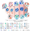
The spleen is a hub of activity during Plasmodium infection. The individual panels display the different structural features of the murine spleen and how they change over the course of a blood‐stage infection with Plasmodium as denoted by the timeline under the spleen. (A) The white pulp is organized into distinct zones: T cell, B cell, and marginal, in the naïve spleen. Inset 1 denotes the diverse array and organization of macrophage populations in the marginal zone and red pulp and the marginal zone B cells. The red arrow denotes the flow of blood out of an arteriole in this region. Also highlighted is the marginal zone bridging channel where the T cell zone abuts the red pulp. These channels serve as an exit site for lymphocytes to move from the white into the red pulp. (B) During the early stages of the infection, polyclonal activation of B cells leads to their expansion and differentiation into Ab‐secreting plasmablasts within the red pulp. Inset 2 highlights the differentiation of inflammatory monocytes recruited from the bone marrow or derived from atypical myeloid precursors in the spleen into macrophages that will aid red pulp macrophages in the phagocytosis and elimination of pRBCs. Inset 3 highlights the atypical progenitor B cells that develop into mature B cells in the spleen during infection with the assistance of CXCL12‐producing FRCs. Production of IL‐17 by the progenitor B cells stimulates CXCL12 production by the FRCs surrounding the white pulp. (C) Activation of CD4+ T cells in the white pulp leads to their differentiation into effector phenotypes. Inset 4 displays these early activation events and the emergence of an intermediate T cell with a mixed Th1/Tfh‐like phenotype before commitment of the cell toward a Th1 or Tfh cell phenotype. Inset 5 displays migration of CD4+ T cells to the border of the T:B cell zones where their interaction with Ag‐specific B cells, specifically ICOS:ICOSL interactions, facilitates their entry into the B cell follicle and promotes their commitment to a GC Tfh cell phenotype. (D) During the later stages of the infection, the GC structure takes shape highlighted by distinct dark (DZ) and light (LZ) zones. While Ab‐secreting cells derived from outside the GC are present at this time, those derived from the GC begin to emerge. Different functional subsets of CD4+ T cells become more prevalent at this time. Inset 6 indicates the movement of Th1 effector cells toward a T regulatory type 1 (Tr1) phenotype that serves to limit the Th1 and Tfh effector response through coproduction of IFN‐γ and IL‐10. Inset 7 shows the role that Tfh cells play in the GC response, serving to promote affinity maturation of GC B cells in the LZ that have undergone somatic mutation of their B cell receptors. (E) Following resolution of the infection, the spleen significantly contracts in size. The spleen color changes from a red to a darker reddish‐brown due to the hemozoin that accumulates in this tissue, primarily found inside macrophages. The GCs contract in size, and the marginal zone becomes reorganized again. Inset 8 shows the emergence of long‐lived plasma cells and memory B cells from the GC. Memory B cells exit this organ via the blood to circulate through lymphoid tissue surveying for Ag. In comparison, plasma cells travel to the bone marrow via the blood to seek a niche for survival and long‐term Ab production. Abs produced by the plasma cells that emerge from the GC will help facilitate the clearance of the pRBCs by macrophages following opsonization. Inset 9 displays the distribution of key memory T cell populations. IL‐7R+CD62Lhi TCM cells are found in circulation and lymphoid tissues and express markers that allow them to traffic through lymphoid tissue. IL‐7RCD62Llo TEM cells will circulate in the blood and possess the ability to enter and exit peripheral tissues to survey for Ag. Individual cell types and key proteins expressed by these cells are highlighted in the key.

References

    1. Steiniger B, Barth P. Microanatomy and function of the spleen. Adv Anat Embryol Cell Biol. 2000;151:III‐IX. 1–101. - PubMed
    1. Beeson JG, Drew DR, Boyle MJ, Feng G, Fowkes FJ, Richards JS. Merozoite surface proteins in red blood cell invasion, immunity and vaccines against malaria. Fems Microbiol Rev. 2016;40:343‐372. - PMC - PubMed
    1. Dorn A, Stoffel R, Matile H, Bubendorf A, Ridley RG. Malarial haemozoin/β‐haematin supports haem polymerization in the absence of protein. Nature. 1995;374:269‐271. - PubMed
    1. Ridley RG, Dorn A, Matile H, Kansy M. Haem polymerization in malaria. Nature. 1995;378:138‐139. - PubMed
    1. Egan TJ, Ross DC, Adams PA. Quinoline anti‐malarial drugs inhibit spontaneous formation of β‐haematin (malaria pigment). FEBS Lett. 1994;352:54‐57. - PubMed

Publication types