. 2021 Mar 15;131(6):e138267.
doi: 10.1172/JCI138267.

Impaired complex I repair causes recessive Leber's hereditary optic neuropathy

Affiliations

Impaired complex I repair causes recessive Leber's hereditary optic neuropathy

Sarah L Stenton et al. J Clin Invest. .

Abstract

Leber's hereditary optic neuropathy (LHON) is the most frequent mitochondrial disease and was the first to be genetically defined by a point mutation in mitochondrial DNA (mtDNA). A molecular diagnosis is achieved in up to 95% of cases, the vast majority of which are accounted for by 3 mutations within mitochondrial complex I subunit-encoding genes in the mtDNA (mtLHON). Here, we resolve the enigma of LHON in the absence of pathogenic mtDNA mutations. We describe biallelic mutations in a nuclear encoded gene, DNAJC30, in 33 unsolved patients from 29 families and establish an autosomal recessive mode of inheritance for LHON (arLHON), which to date has been a prime example of a maternally inherited disorder. Remarkably, all hallmarks of mtLHON were recapitulated, including incomplete penetrance, male predominance, and significant idebenone responsivity. Moreover, by tracking protein turnover in patient-derived cell lines and a DNAJC30-knockout cellular model, we measured reduced turnover of specific complex I N-module subunits and a resultant impairment of complex I function. These results demonstrate that DNAJC30 is a chaperone protein needed for the efficient exchange of complex I subunits exposed to reactive oxygen species and integral to a mitochondrial complex I repair mechanism, thereby providing the first example to our knowledge of a disease resulting from impaired exchange of assembled respiratory chain subunits.

Keywords: Genetic diseases; Genetics; Neuroscience.


Conflict of interest statement

Conflict of interest: CBC, CK, PB, CLM, TK, and VC have received research support, speaker honoraria, consulting fees, and travel reimbursement from Santhera Pharmaceuticals and GenSight Biologics.

Figures

Figure 1
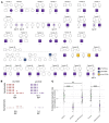
Figure 1. Identification of pathogenic DNAJC30 mutations in LHON patients in association with a complex I defect.
(A) Pedigrees from 29 families. The genotype is denoted by (–/–) for homozygous variant carriers, (+/–) for heterozygous variant carriers, and (+/+) for carriers of 2 wild-type alleles. Individuals with a central dot are homozygous carriers (–/–) who did not express the disease phenotype at their current age, stated beneath. (B) Schematic of the sex-dependent incomplete penetrance in both maternally inherited LHON (mtLHON) and recessive LHON (arLHON), demonstrating a clear male predominance in symptomatic carriers of disease-causing variants. (C) Mitochondrial complex I–dependent (CI-dependent) respiration rate measurement in control (n = 30, technical replicates) and arLHON (n = 28, technical replicates) fibroblast cell lines, demonstrating a mild respiratory defect rescued by reexpression of naive DNAJC30 (arLHON-rescue, n = 36, technical replicates). The defect in CI-dependent respiration rate is recapitulated in the DNAJC30-KO (n = 25, technical replicates) in comparison with control (n = 43, technical replicates) HEK293 cell lines. Data are normalized to the respective control cell line and depicted by the mean ± SD; 2-sided Student’s t test, P values corrected for multiple comparisons to the control (Dunnett’s test). ****P ≤ 0.0001. NS, not significant.
Figure 2
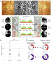
Figure 2. LHON associated with DNAJC30 mutations presents as a phenocopy of maternally inherited LHON.
(A) The pathognomonic triad of ophthalmological features in mtLHON is recapitulated in arLHON. Presented here is an illustrative example from 1 arLHON patient. Top panel: Optic nerve head picture and fluorescein angiography in the acute stage of the disease displaying microangiopathy without leakage, fiber swelling, and initial temporal pallor of the optic disc. Bottom panel: The retinal nerve fiber layer (RNFL) thickness analysis and deviation map showing the progressive thinning of fibers from the subacute stage (right eye [OD] at 2 months and left eye [OS] at 3 months after visual loss) to the chronic stage (3 years). The 30° Humphrey visual field shows progressive enlargement of the central scotoma in the subacute stage (from 2 to 4 months in OD and from 3 to 5 months in OS) and fenestration of the scotoma after 3 years (10° Humphrey visual field) associated with recovery of visual acuity (VA, expressed in decimal units). m, months; y, years. RNFL thickness (middle) is displayed as a function of the quadrant in the deviation map: temporal (TEMP), superior (SUP), nasal (NAS), and inferior (INF). (B) Age of onset (years) in mtLHON (n = 104) (28) and arLHON (n = 31). Data presented as mean ± SD. ****P ≤ 0.0001 by 2-sided Student’s t test. (C) Spontaneous and idebenone-treated rate of clinically significant recovery of VA, defined as improvement in logMAR VA of ≥0.2, in mtLHON (4, 5, 6) in comparison with arLHON, in which treated recovery rates were significantly higher in arLHON (mtLHON 43.6%, arLHON 80.6%, P < 0.001, Fisher’s exact test).
Figure 3
Figure 3. DNAJC30 mutations result in impaired repair of specific subunits of mitochondrial complex I.
(A) The structure of mitochondrial complex I (CI) (30), depicted by module and respective protein. (B) Mitochondrial CI structure colored by the mean degree of subunit turnover in 12 hours in control fibroblast cell lines (n = 7) and patient fibroblast cell lines (n = 6) depicted as a percentage. The mean data are provided in Supplemental Table 15 and the individual experiments are depicted in Supplemental Tables 11–14. (C) The DNAJC30 interacting partners in CI according to the BioPlex database highlighted on the CI structure. The interaction partners in the N-module (NDUFV3, NDUFS4, NDUFS6, and NDUFA7) account for 4 of the 5 CIHIGH subunits, defined as subunits with >25% turnover in 12 hours in the control fibroblast cell lines. (D) Turnover measurement of CIHIGH subunits (n = 5) and (E) CIMOD subunits (n = 5) in 12 hours in control (n = 7), arLHON (n = 6), and mtLHON patient (n = 3, m.3460G>A in MT-ND1, m.11778G>A in MT-ND4, and m.14484T>C in MT-ND6) fibroblast cell lines, and control (n = 1) and DNAJC30-KO (n = 1) HEK293 cell lines. arLHON patients demonstrate a defect in CIHIGH (control mean 33.6% ± 11.2% SD, patient mean 16.8% ± 5.5% SD) and CIMOD (control mean 18.3% ± 5.7% SD, patient mean 12.5% ± 3.9% SD) subunits. The defective turnover of CIHIGH subunits is shown to be specific to arLHON (CIHIGH subunits, control mean 33.6% ± 11.2% SD, mtLHON mean 33.7% ± 13.7% SD). The DNAJC30-KO HEK293 cell line demonstrates a defect in CIHIGH (control mean 48.7% ± 8.3% SD, KO mean 31.0% ± 11.6% SD) and CIMOD (control mean 36.6% ± 8.9% SD, KO mean 24.5% ± 7.5% SD) subunits. Data depicted as the mean ± SD; 2-sided Student’s t test, P values corrected for multiple comparisons to the control (Dunnett’s test). *P ≤ 0.05, **P ≤ 0.01, ***P ≤ 0.001. NS, not significant. A complete summary of the data is provided in Supplemental Table 10 and the experiment is depicted in Supplemental Tables 10–15 and 17.
Figure 4
Figure 4. Schematic representation of the proposed role of DNAJC30 in complex I repair.
(A) In normal physiological conditions, DNAJC30 interacts with specific complex I (CI) N-module proteins (CIHIGH), facilitating their disassembly and subsequent degradation. In the setting of highly functional CI, these proteins are newly synthesized and replaced without degradation of further subunits. In the case of oxidative damage to the CI N-module, upon disassembly of the CIHIGH subunits by DNAJC30 the protease CLPXP may access and remove the damaged CIMOD subunits (20, 21). Along with the CIHIGH subunits, these subunits are subsequently resynthesized and replaced, negating the need for complete degradation and synthesis of CI at high energetic cost. (B) Compared with control, in the presence of DNAJC30 mutations turnover of the N-module subunits is decreased, impairing the CI repair mechanism and leading to the accumulation of CI with reduced function.

Comment in

References

    1. Von Graefe A. Exceptionelles verrhalten des gesichtsfeldes bei pigmentenartung der netzhaut. Albrecht Von Graefes Arch Ophthalmol. 1858;4(6):250–253. - PubMed
    1. Leber TH. Ueber hereditäre und congenital-angelegte Sehnervenleiden. Albrecht Von Graefes Arch Ophthalmol. 1871;17(2):249–291. doi: 10.1007/BF01694557. - DOI
    1. Yu-Wai-Man P, et al. A neurodegenerative perspective on mitochondrial optic neuropathies. Acta Neuropathol. 2016;132(6):789–806. doi: 10.1007/s00401-016-1625-2. - DOI - PMC - PubMed
    1. Catarino CB, et al. Real-world clinical experience with idebenone in the treatment of leber hereditary optic neuropathy. J Neuroophthalmol. 2020;40(4):558–565. doi: 10.1097/WNO.0000000000001023. - DOI - PMC - PubMed
    1. Carelli V, et al. Idebenone treatment in Leber’s hereditary optic neuropathy. Brain. 2011;134(9):e188–e188. doi: 10.1093/brain/awr180. - DOI - PubMed

Publication types

MeSH terms