. 2021 Jan 19;16(1):e0245555.
doi: 10.1371/journal.pone.0245555. eCollection 2021.

A versatile cryo-transfer system, connecting cryogenic focused ion beam sample preparation to atom probe microscopy

Affiliations

A versatile cryo-transfer system, connecting cryogenic focused ion beam sample preparation to atom probe microscopy

Chandra Macauley et al. PLoS One. .

Abstract

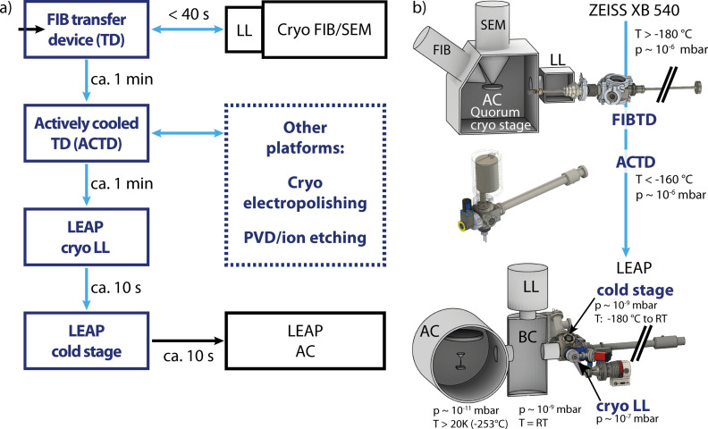
Atom probe tomography (APT) is a powerful technique to obtain 3D chemical and structural information, however the 'standard' atom probe experimental workflow involves transfer of specimens at ambient conditions. The ability to transfer air- or thermally-sensitive samples between instruments while maintaining environmental control is critical to prevent chemical or morphological changes prior to analysis for a variety of interesting sample materials. In this article, we describe a versatile transfer system that enables cryogenic- or room-temperature transfer of specimens in vacuum or atmospheric conditions between sample preparation stations, a focused ion beam system (Zeiss Crossbeam 540) and a widely used commercial atom probe system (CAMECA LEAP 4000X HR). As an example for the use of this transfer system, we present atom probe data of gallium- (Ga)-free grain boundaries in an aluminum (Al) alloy specimen prepared with a Ga-based FIB.


Conflict of interest statement

The authors have declared that no competing interests exist.

Figures

Fig 1
Fig 1. An experimental overview of the transfer system.
In a) the workflow is shown with transfer times. Custom components designed and built as part of this work are blue while existing systems are black A schematic overview in b) shows the FIB transfer device connected directly to a Zeiss Crossbeam 540 FIB/SEM. Prepared samples can be transferred with an actively cooled transfer device (ACTD) that interfaces with existing instrument load locks (LL) into a CAMECA LEAP. AC: analysis chamber, BC: buffer chamber, RT: room temperature.
Fig 2
Fig 2. The cryo stage and FIB transfer device (FIBTD) enable characterization, sample preparation and transfer at cryogenic conditions.
a) A custom 54° pre-tilted FIB cryo-shuttle mounted on the Quorum cryo-stage with key components and directions labeled. b) Rendered CAD drawing of the FIBTD indicating the flexible bellows coupling, 1, and the two possible connection points for the actively cooled transfer device (ACTD). c) The FIBTD connects directly to the existing FIB load-lock. d) The non-pre-tilted (top) and 54° pre-tilted FIB cryo-shuttles (bottom) attach to the end of the FIBTD with a bayonet connection and are thermally-isolated from the transfer rod with a polyether ether ketone end-component, 2.
Fig 3
Fig 3. The actively cooled transfer device (ACTD) and the double nipple sample shuttle enable transfers to the atom probe.
a) Photo of the ACTD showing the valve open and the copper end-component (labeled 1) extended. b) The rendered cross-section of the ACTD shows the threaded connection between the liquid nitrogen (LN2) cold finger and the copper end-component (1). A low thermal conductivity PEEK component (2) thermally isolates the end-component from the rest of the transfer rod. Temperature is measured at the cold finger using a thermocouple (TC). c) A higher magnification inset of b) more clearly shows how the double nipple screws into the copper end-component during transfers, resulting in the atom probe tip being shielded. d) Transfers between the FIBTD and ACTD are enabled by the two-threaded double nipple that has a right-handed screw (RHS) thread and a left-handed screw (LHS) thread.
Fig 4
Fig 4. Samples are handed-off between the FIBTD and the ACTD while the FIBTD is connected to the FIB load lock (LL).
a) CAD drawing depicting the 54° connection between the FIBTD and the ACTD. The perpendicular port for the ACTD that is labeled ACTD 2 can be used with the FIB cryo shuttles that are not pre-tilted. b) The connected transfer devices interface directly to the Zeiss load lock. When the FIB cryo shuttle is first removed from the FIB, c), it must be rotated 90° along the axis of the FIBTD transfer rod to line-up with the ACTD as shown in d). The copper end-component of the ACTD then surrounds the sample, e) as the double nipple is passed from the FIBTD to the ACTD.
Fig 5
Fig 5. Modifications were made to a LEAP to enable cryo/environmental transfers.
a) The rendered CAD drawing indicates the modifications to the LEAP, described in the text. A detailed view of the cryogenic-stage, is shown in b) with a custom LEAP shuttle in place as it would be during the hand-off of the sample and double nipple from the ACTD. The region highlighted with an orange box in a) is shown in c) to illustrate the movement of the cryo-stage from the perspective of the analysis chamber. d) A photo shows in detail the connection of the ACTD to the LEAP.
Fig 6
Fig 6. Results of cryo-preparation of nanocrystalline-Al.
a) microstructure and resulting sample in the LEAP, showing no frost formation. b) overview of the reconstructed dataset. The red box corresponds to the data shown in c) detail of grain boundary showing change in crystallography and distribution of Ga. No segregation of Ga is visible at the grain boundary. d) Concentration profile of Ga across the grain boundary depicted in c). e) Concentration of Ga along the entire dataset, exhibiting a typical implantation profileNo enrichment at the grain boundary is observed.

References

    1. Dubochet J. Cryo-EM-the first thirty years. J Microsc 2012;245:221–4. 10.1111/j.1365-2818.2011.03569.x - DOI - PubMed
    1. Henderson R, Baldwin JM, Ceska TA, Zemlin F, Beckmann E, Downing KH. Model for the structure of bacteriorhodopsin based on high-resolution electron cryo-microscopy. J Mol Biol 1990;213:899–929. 10.1016/S0022-2836(05)80271-2 - DOI - PubMed
    1. Li Y, Li Y, Pei A, Yan K, Sun Y, Wu C-L, et al. Atomic structure of sensitive battery materials and interfaces revealed by cryo–electron microscopy. Science (80-) 2017;358:506–10. 10.1126/science.aam6014 - DOI - PubMed
    1. Hassan AN, Frank JF, Elsoda M. Observation of bacterial exopolysaccharide in dairy products using cryo-scanning electron microscopy. Int Dairy J 2003;13:755–62. 10.1016/S0958-6946(03)00101-8 - DOI
    1. Schertel A, Snaidero N, Han HM, Ruhwedel T, Laue M, Grabenbauer M, et al. Cryo FIB-SEM: Volume imaging of cellular ultrastructure in native frozen specimens. J Struct Biol 2013;184:355–60. 10.1016/j.jsb.2013.09.024 - DOI - PubMed

Publication types