. 2021 Jan 19;16(1):e0245239.
doi: 10.1371/journal.pone.0245239. eCollection 2021.

Hmx1 regulates urfh1 expression in the craniofacial region in zebrafish

Affiliations

Hmx1 regulates urfh1 expression in the craniofacial region in zebrafish

Younes El Fersioui et al. PLoS One. .

Abstract

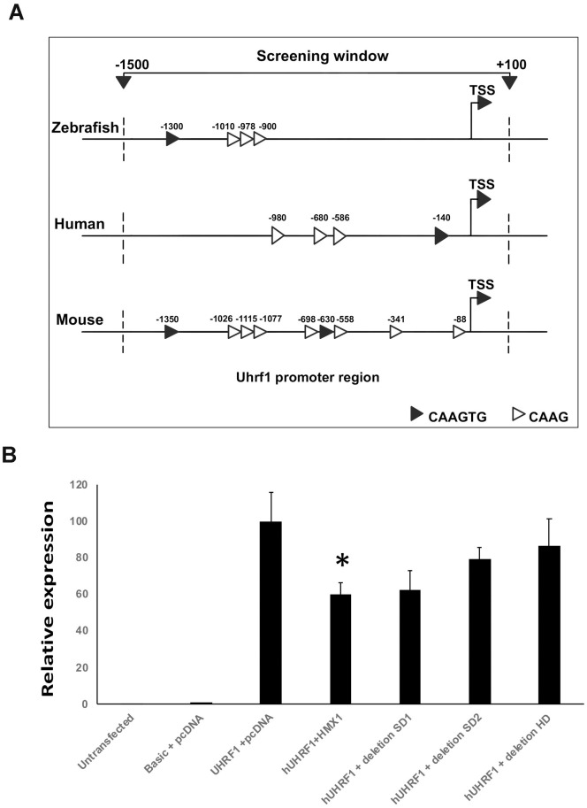
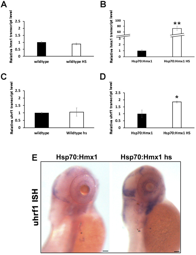
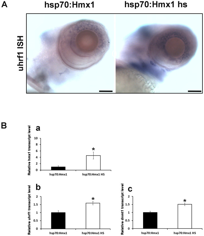
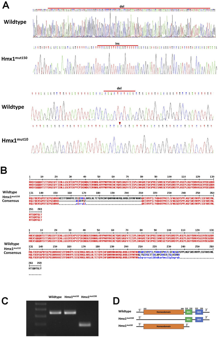
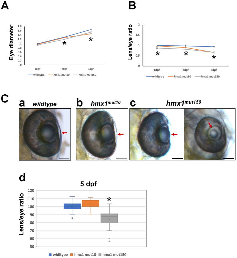
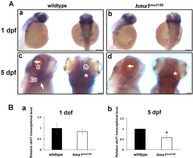
H6 family homeobox 1 (HMX1) regulates multiple aspects of craniofacial development as it is widely expressed in the eye, peripheral ganglia and branchial arches. Mutations in HMX1 are linked to an ocular defect termed Oculo-auricular syndrome of Schorderet-Munier-Franceschetti (MIM #612109). We identified UHRF1 as a target of HMX1 during development. UHRF1 and its partner proteins actively regulate chromatin modifications and cellular proliferation. Luciferase assays and in situ hybridization analyses showed that HMX1 exerts a transcriptional inhibitory effect on UHRF1 and a modification of its expression pattern. Overexpression of hmx1 in hsp70-hmx1 zebrafish increased uhrf1 expression in the cranial region, while mutations in the hmx1 dimerization domains reduced uhrf1 expression. Moreover, the expression level of uhrf1 and its partner dnmt1 was increased in the eye field in response to hmx1 overexpression. These results indicate that hmx1 regulates uhrf1 expression and, potentially through regulating the expression of factors involved in DNA methylation, contribute to the development of the craniofacial region of zebrafish.


Conflict of interest statement

The authors have declared that no competing interests exist.

Figures

Fig 1
Fig 1
Schematic representation of the HMX1 predicted promoter region in zebrafish, human and mouse genomes (A). Note the presence of complete Hmx1 binding sites (CAAGTG) and the minimal binding core (CAAG). Activity of HMX1 on human UHRF1 promoter (B). The pGL3-Uhrf1 reporter plasmid was cotransfected with pcDNA3.1 empty, pcDNA3.1-Hmx1 expression vector, and pcDNA3.1-Hmx1 del SD1, del SD2, or del HD. HMX1 inhibits UHRF1 transcriptional activity by 40%. HMX1 del SD1, HMX1 del SD2 and HMX1 del HD had no statistical significant effect. Luciferase activity was normalized against β-galactosidase activity. Data are expressed as mean of three or more experiments. *; P<0.05 (ANOVA test).
Fig 2
Fig 2. Expression of Uhrf1 in hsp70:Hmx1 transgenic zebrafish at 5 dpf.
(A-D) Quantitative RT-PCR from RNA isolated from zebrafish at 5 dpf. (A) Expression of hmx1 in wildtype zebrafish without or with heat shock. (B) Expression of hmx1 in transgenic zebrafish without or with heat shock. (C) Expression of uhrf1 in wildtype zebrafish without or with heat shock. (D) Expression of uhrf1 in transgenic zebrafish without or with heat shock. Heat shock treatment induced expression of Hmx1 in hsp70:Hmx1 but not in wildtype zebrafish. Uhrf1 expression increased in hsp70:Hmx1 zebrafish embryos at 5dpf. (E) Lateral view of hsp70:Hmx1 zebrafish embryos at 5 dpf. In situ hybridization was performed with antisense RNA probe specific for uhrf1. Uhrf1 expression increased in response to ubiquitous hmx1 heat activation, but remained limited to the dorsal, eye and branchial arches regions. Bar, E 100 μm.
Fig 3
Fig 3. Lateral view of cranial region of hsp70:Hmx1 zebrafish embryos at 5 dpf.
(A) In situ hybridization specific for uhrf1. Hmx1 heatshock experiments induced uhrf1 expression in the retina and branchial arches region. Quantification of RNA transcript isolated from zebrafish retinas at 5 dpf. (B) Uhrf1 (b) and dnmt1 (c) expression increased in response to the activation of hmx1 in hsp70:Hmx1 embryos. Bar, A 75 μm.
Fig 4
Fig 4. Hmx1 mutant zebrafish generation via zinc-finger nuclease method.
(A) Wildtype and hmx1 mutant sequence analysis. Hmx1mut150 mutant embryos carried a 170 bp deletion and a 21 bp insertion, while Hmx1mut10 embryos reported a 10 bp deletion. (B,D) ExPasy translation tool depicted the disruption of the encoding sequence of the homeodomain and the downstream region composed of SD1 domains in exon2. C. Agarose gel of PCR amplified DNA products. The DNA isolated from WT, hmx1mut10 and hmx1mut150 embryos was amplified by PCR.
Fig 5
Fig 5. Morphometric analysis of the developing zebrafish eye at early stages.
(A) Tracking of the growth of wildtype, hmx1mut10 and hmx1mut150 eye from 1 dpf to 3 dpf. Hmx1mut10 and hmx1mut150 eyes are reduced in size compared to wildtype embryos at 2 and 3 dpf stages. (B) Lens growth expressed as ratio lens/whole eye. hmx1mut10 and hmx1mut150 lenses are smaller at 3 dpf. (C) Wildtype, hmx1mut10 and hmx1mut150 eyes at 5 dpf. Wildtype and hmx1mut10 present a fully developed lens (5C, a-b), while hmx1mut150 embryos exhibit two opposing phenotypes (5C, c); on the left side a recovered lens is presented, while on the right a smaller lens presenting a clear phenotype called microphakia. (5C, d) graphic representation of lens/eye ratio; hmx1mut150 presents a wide range of distribution of the lens size. Red arrows indicate the lens at 5 dpf. Data are expressed as mean of three to 5 experiments. *; P<0.05 (ANOVA test). Bar, C 100 μm.
Fig 6
Fig 6. In situ hybridization specific for uhrf1 in wildtype and hmx1 mutant zebrafish at 1 dpf.
(A) uhrf1 is expressed in the dorsal region and in the eye field region at 1 dpf in both wildtype and hmx1 mutant embryos (a,b). c,d: uhrf1 expression is reduced in the eye field, in the forebrain and branchial region (c,d). White arrow, retina and branchial arches; asterisk, forebrain. e, eye; fb, forebrain; ba, branchial arches. RT-PCR quantification of uhrf1 transcript isolated from the cranial region. (B) Uhrf1 expression is reduced at 5 dpf (b) but not at 24 hpf (a). Bar, A 100 μm.

Similar articles

Cited by

References

    1. Mayor R, Theveneau E. The neural crest. Development. 2013;140(11):2247–51. Epub 2013/05/16. 10.1242/dev.091751 . - DOI - PubMed
    1. Williams AL, Bohnsack BL. Neural crest derivatives in ocular development: discerning the eye of the storm. Birth Defects Res C Embryo Today. 2015;105(2):87–95. Epub 2015/06/06. 10.1002/bdrc.21095 . - DOI - PMC - PubMed
    1. Trainor PA, Krumlauf R. Hox genes, neural crest cells and branchial arch patterning. Curr Opin Cell Biol. 2001;13(6):698–705. Epub 2001/11/08. 10.1016/s0955-0674(00)00273-8 . - DOI - PubMed
    1. Meinecke L, Sharma PP, Du H, Zhang L, Nie Q, Schilling TF. Modeling craniofacial development reveals spatiotemporal constraints on robust patterning of the mandibular arch. PLoS Comput Biol. 2018;14(11):e1006569 Epub 2018/11/28. 10.1371/journal.pcbi.1006569 . - DOI - PMC - PubMed
    1. Fadool JM, Dowling JE. Zebrafish: a model system for the study of eye genetics. Prog Retin Eye Res. 2008;27(1):89–110. Epub 2007/10/27. 10.1016/j.preteyeres.2007.08.002 . - DOI - PMC - PubMed

Publication types

MeSH terms

LinkOut - more resources