. 2021 Jan 19;16(1):e0245541.
doi: 10.1371/journal.pone.0245541. eCollection 2021.

The role of posttranslational modifications in generating neo-epitopes that bind to rheumatoid arthritis-associated HLA-DR alleles and promote autoimmune T cell responses

Affiliations

The role of posttranslational modifications in generating neo-epitopes that bind to rheumatoid arthritis-associated HLA-DR alleles and promote autoimmune T cell responses

Stephane Becart et al. PLoS One. .

Abstract

While antibodies to citrullinated proteins have become a diagnostic hallmark in rheumatoid arthritis (RA), we still do not understand how the autoimmune T cell response is influenced by these citrullinated proteins. To investigate the role of citrullinated antigens in HLA-DR1- and DR4-restricted T cell responses, we utilized mouse models that express these MHC-II alleles to determine the relationship between citrullinated peptide affinity for these DR molecules and the ability of these peptides to induce a T cell response. Using a set of peptides from proteins thought to be targeted by the autoimmune T cell responses in RA, aggrecan, vimentin, fibrinogen, and type II collagen, we found that while citrullination can enhance the binding affinity for these DR alleles, it does not always do so, even when in the critical P4 position. Moreover, if peptide citrullination does enhance HLA-DR binding affinity, it does not necessarily predict the generation of a T cell response. Conversely, citrullinated peptides can stimulate T cells without changing the peptide binding affinity for HLA-DR1 or DR4. Furthermore, citrullination of an autoantigen, type II collagen, which enhances binding affinity to HLA-DR1 did not enhance the severity of autoimmune arthritis in HLA-DR1 transgenic mice. Additional analysis of clonal T cell populations stimulated by these peptides indicated cross recognition of citrullinated and wild type peptides can occur in some instances, while in others cases the citrullination generates a novel T cell epitope. Finally, cytokine profiles of the wild type and citrullinated peptide stimulated T cells unveiled a significant disconnect between proliferation and cytokine production. Altogether, these data demonstrate the lack of support for a simplified model with universal correlation between affinity for HLA-DR alleles, immunogenicity and arthritogenicity of citrullinated peptides. Additionally they highlight the complexity of both T cell receptor recognition of citrulline as well as its potential conformational effects on the peptide:HLA-DR complex as recognized by a self-reactive cell receptor.


Conflict of interest statement

This work was supported by a Merit Review grant from the Department of Veterans Affairs (E.F.R.), and by funding from Janssen Research and Development (E.F.R.). Janssen Research and Development provided financial support in the form of authors’ salaries (S.B and N.L.R.), study design, data analyses, and preparation of the manuscript. This commercial affiliation does not alter our adherence to all PLOS ONE policies on sharing data and materials.

Figures

Fig 1
Fig 1. Measurement of peptide binding to DRB1*01:01.
10 nM of soluble DR1 was incubated for 18 h at 37° C with 0.5 nM of biotinylated CII(257–274) peptide and various concentrations of WT and Cit-peptides as competitors. Bound peptides were separated from free peptides by immobilizing the DR molecules with an antibody on microtiter plates. Bound biotinylated peptide was detected by incubation with streptavidin-europium. Time resolved fluorescence was quantitated and data are expressed as relative fluorescence units measured. Each binding assay was performed in duplicate, and data are representative of 3 experiments.
Fig 2
Fig 2. Measurement of peptide binding to DRB1*04:01.
10 nM of soluble DR4 was incubated for 18 h at 37° C with 0.5 nM of biotinylated CII(257–274) peptide and various concentrations of WT and Cit-peptides as competitors. Bound peptides were separated from free peptides by immobilizing the DR molecules with an antibody on microtiter plates. Bound biotinylated peptide was detected by incubation with streptavidin-europium. Time resolved fluorescence was quantitated and data are expressed as relative fluorescence units measured. Each binding assay was performed in duplicate, and data are representative of 3 experiments.
Fig 3
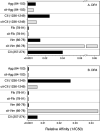
Fig 3. Relative affinity of WT and Cit-peptide binding to DRB1*01:01 and DRB1*04:01.
The 50% inhibitory concentration values (IC50) values were calculated from the binding curves in Figs 1 and 2, and plotted as 1/IC50 to indicate relative affinity. Panel A, DR1 binding; panel B, DR4 binding.
Fig 4
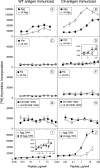
Fig 4. Immunogenicity and antigenicity of WT and Cit-peptides in DRB1*01:01 and DRB1*04:01 humanized mice.
B6.DR1 and B6.DR4 mice were immunized with the WT and Cit-peptides, and 10 days later, T cells from draining lymph nodes were cultured in the presence of various concentrations of peptides for 4 days. On day 3, [3H]-thymidine was added to the cultures and cells were harvested the following day. Data in the panels on the left were derived from WT peptide immunized mice; data on the right were from the corresponding Cit-peptide immunizations. Panels A thru H are from B6.DR1 mouse immunizations; panels I and J are from B6.DR4 immunized mice. Controls were unstimulated cells, and data shown are Δ cpm (experimental minus control). Error bars indicate standard deviation of triplicates, and data are representative of two independent experiments.
Fig 5
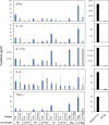
Fig 5. Cytokine production by T cells stimulated with WT or Cit-peptides.
Supernatants from T cell proliferation assays in Fig 4 were collected 3 days after antigen stimulation and analyzed for the expression of proinflammatory cytokines. Cytokines produced were measured using a multiplexed bead assay and analyzed using a BioRad MagPix. Standard curves were used to calculate cytokine concentration. The cytokines produced by CII immunized T cells stimulated with the immunodominant epitope, CII(257–274), that drives autoimmune arthritis in the B6.DR1 model are shown for comparison (right panels). Data are representative of two independent experiments and based on duplicate samples.
Fig 6
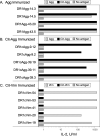
Fig 6. T cell receptor cross recognition of WT and Cit-peptides.
Clonal populations of T cells were tested for their ability to recognize WT or Cit-peptides in antigen presentation assays as described in Materials and Methods. T cells from Agg, Cit-Agg, and Cit-Vim immunized mice were used to produce T-cell hybridomas, and multiple clones from each were tested for their ability to recognize the immunogen and the corresponding WT or Cit-peptide. Data indicate that cross recognition of WT and Cit-peptides can occur at the clonal level, and that Cit-peptides can be novel antigens for T cell responses. Data shown are representative responses from multiple clones tested from each immune T cell population.
Fig 7
Fig 7. Measurement of PAD4-meidated citrullination of CII.
Native bovine CII was citrullinated using PAD4, and the degree of citrullination achieved was measured using a colorimetric assay and absorbance at 530 nm. Cit-CII peptide was produced synthetically and used for comparison. All samples were dissolved in 10 mM acetic, and this reagent was used as the negative control. Data represent triplicate measurements, and error bars represent standard deviation.
Fig 8
Fig 8. Induction of autoimmune arthritis in HLA-DR1 mice with WT CII and Cit-CII.
B6.DR1 mice were immunized with native CII or Cit-CII and the incidence (panel A) and severity (panels B, C, and D) of arthritis were monitored. Disease severity was evaluated on the basis of arthritic animals only, and was assessed using a scale of 0 to 4 as described in Materials and Methods. No significant differences were observed in the incidence or severity of disease between the two groups. Data are representative of two independent experiments.

References

    1. Stastny P, Ball EJ, Dry PJ, Nunez G. The human immune response region (HLA-D) and disease susceptibility. Immunol Rev. 1983;70:113–54. 10.1111/j.1600-065x.1983.tb00712.x - DOI - PubMed
    1. du Montcel ST, Michou L, Petit-Teixeira E, Osorio J, Lemaire I, Lasbleiz S, et al. New classification of HLA-DRB1 alleles supports the shared epitope hypothesis of rheumatoid arthritis susceptibility. Arthritis Rheum. 2005;52(4):1063–8. 10.1002/art.20989 - DOI - PubMed
    1. Morgan AW, Haroon-Rashid L, Martin SG, Gooi HC, Worthington J, Thomson W, et al. The shared epitope hypothesis in rheumatoid arthritis: evaluation of alternative classification criteria in a large UK Caucasian cohort. Arthritis Rheum. 2008;58(5):1275–83. 10.1002/art.23432 - DOI - PubMed
    1. Gorga JC, Brown JH, Jardetzky T, Wiley DC, Strominger JL. Crystallization of HLA-DR antigens. Res Immunol. 1991;142(5–6):401–7. 10.1016/0923-2494(91)90038-k - DOI - PubMed
    1. Ferucci ED, Templin DW, Lanier AP. Rheumatoid arthritis in American Indians and Alaska Natives: a review of the literature. Semin Arthritis Rheum. 2005;34(4):662–7. 10.1016/j.semarthrit.2004.08.003 - DOI - PubMed

Publication types