. 2021 Jan 19;16(1):e0245618.
doi: 10.1371/journal.pone.0245618. eCollection 2021.

Myogenin is required for assembly of the transcription machinery on muscle genes during skeletal muscle differentiation

Affiliations

Myogenin is required for assembly of the transcription machinery on muscle genes during skeletal muscle differentiation

Abhinav Adhikari et al. PLoS One. .

Abstract

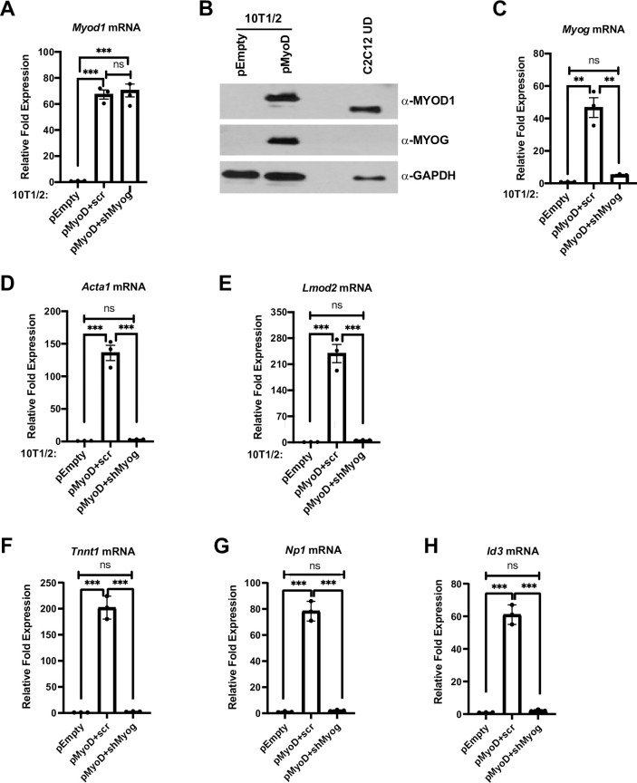
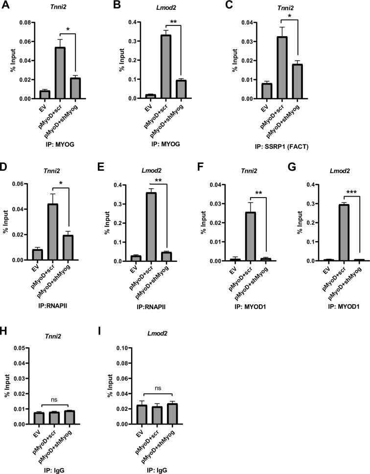
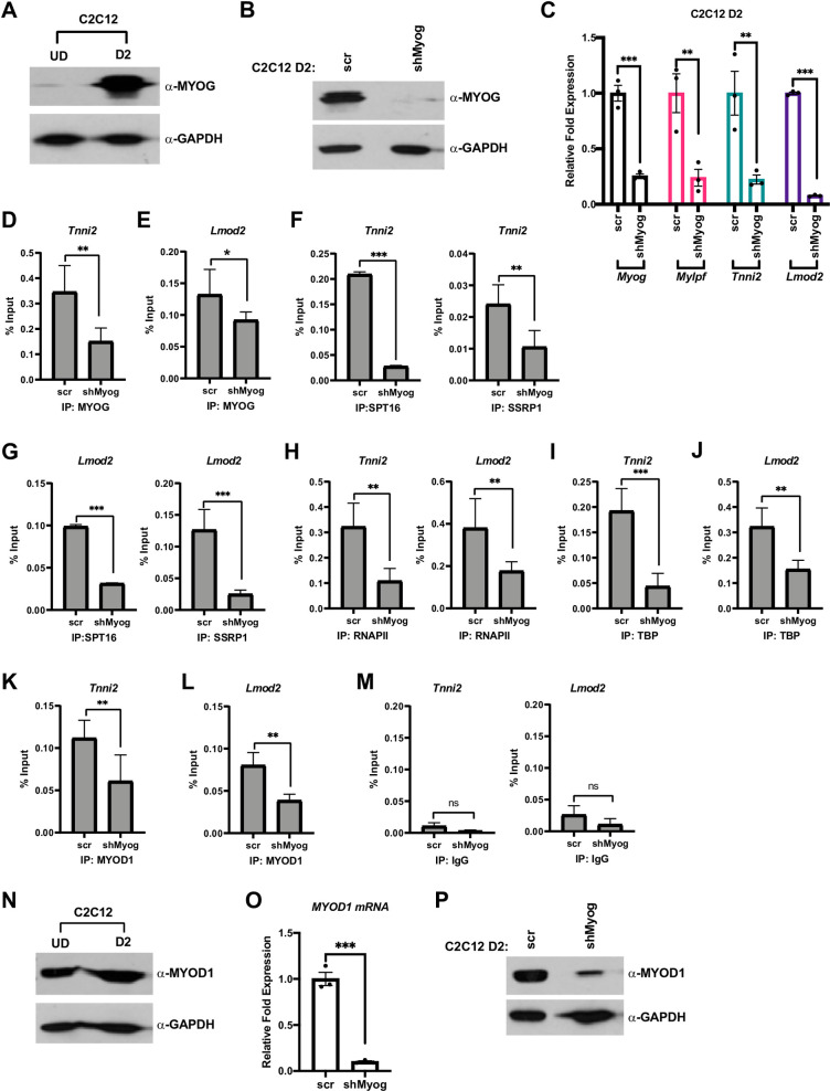
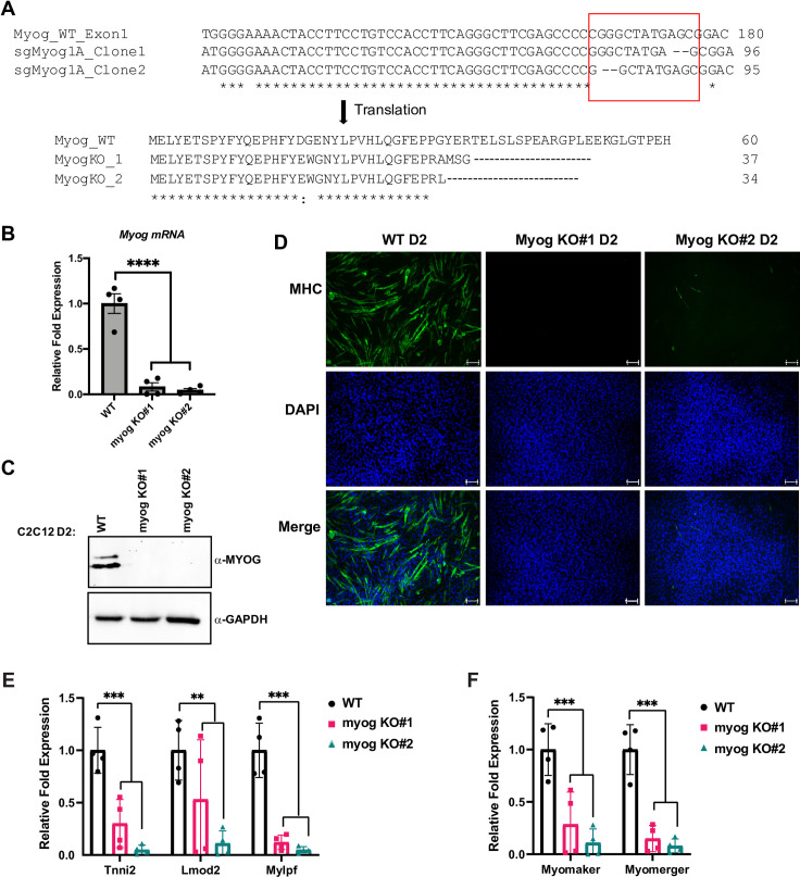
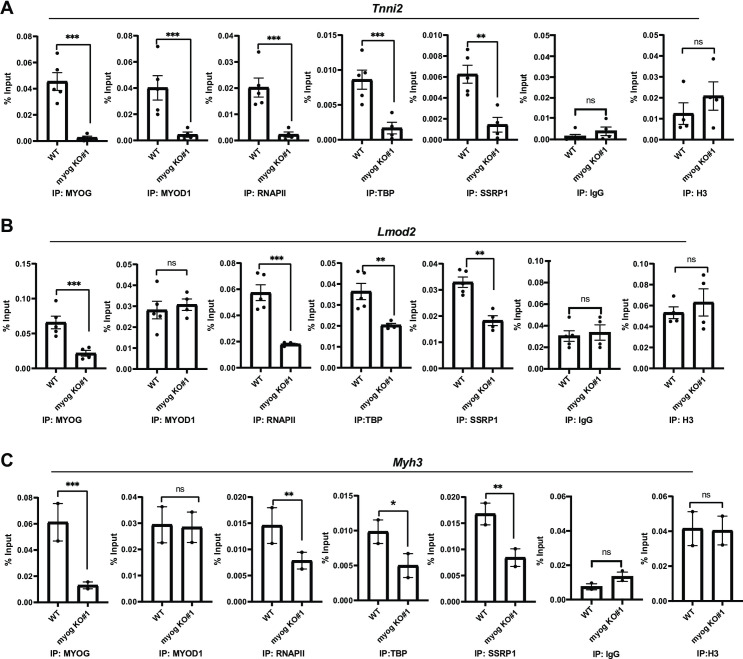
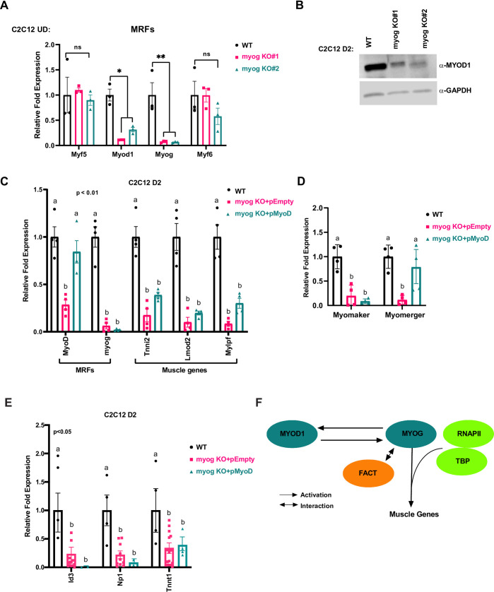
Skeletal muscle gene expression is governed by the myogenic regulatory family (MRF) which includes MyoD (MYOD1) and myogenin (MYOG). MYOD1 and MYOG are known to regulate an overlapping set of muscle genes, but MYOD1 cannot compensate for the absence of MYOG in vivo. In vitro, late muscle genes have been shown to be bound by both factors, but require MYOG for activation. The molecular basis for this requirement was unclear. We show here that MYOG is required for the recruitment of TBP and RNAPII to muscle gene promoters, indicating that MYOG is essential in assembling the transcription machinery. Genes regulated by MYOD1 and MYOG include genes required for muscle fusion, myomaker and myomerger, and we show that myomaker is fully dependent on activation by MYOG. We also sought to determine the role of MYOD1 in MYOG dependent gene activation and unexpectedly found that MYOG is required to maintain Myod1 expression. However, we also found that exogenous MYOD1 was unable to compensate for the loss of Myog and activate muscle gene expression. Thus, our results show that MYOD1 and MYOG act in a feed forward loop to maintain each other's expression and also show that it is MYOG, and not MYOD1, that is required to load TBP and activate gene expression on late muscle gene promoters bound by both factors.


Conflict of interest statement

The authors have declared that no competing interests exists.

Figures

Fig 1
Fig 1. MYOD1 and MYOG are required to induce muscle gene expression in 10T1/2 fibroblast cells.
A-H. Plasmid expressing MYOD1 (pMyoD) or empty (pEmpty) vector were stably transfected in 10T1/2 cells. Cells were harvested for mRNA and protein and were analyzed for Myod1 by qRT-PCR (A), and western blot (B), respectively. C2C12 UD was used as a positive control for MYOD1. GAPDH was used as a loading control. Stable cell lines overexpressing Myod1 (pMyoD) were further stably transfected with shRNA constructs targeting Myog (pMyoD+shMyog) or scrambled control (pMyoD+scr). Total RNA was extracted and was assayed for Myod1 (A), Myog (C), Acta1 (D), Lmod2 (E), Tnnt1 (F), Np1 (G) and Id3 (H). Standard errors (S.E.) from the mean (Mean ± S.E.) represents the error bars. (ANOVA test followed by Tukey’s multiple comparison test; ns represents 'not significant', **p<0.01 and ***p<0.001, n = 3 biological replicates).
Fig 2
Fig 2. Depletion of Myog impairs RNA polymerase II recruitment to muscle genes.
A-G. Stable 10T1/2 cell lines overexpressing Myod1 (pMyoD) were further stably transfected with shRNA constructs targeting Myog (pMyoD+shMyog) or scrambled control (pMyoD+scr). EV represents empty vector (pEF6/V5 His). ChIP assays were performed with antibodies against MYOG (A and B), SSRP1(C), RNAPII (D and E), MYOD1 (F and G) and IgG (H and I) and primers spanning the Tnni2 (A, C, D, F and H) and Lmod2 (B, E, G and I) promoters. Standard errors (S.E.) from the mean (Mean ± S.E.) represents the error bars. (Student t.test; ns represents ‘not significant’, *p<0.05, **p<0.01 and ***p<0.001, n = 3 biological replicates).
Fig 3
Fig 3. MYOG assembles the transcription machinery at muscle gene promoters.
A. C2C12 myoblasts were grown in proliferating condition (U.D.) and differentiated for two days (D2) and were assayed for the expression of MYOG by western blot. GAPDH was used as a loading control. B. C2C12 cells were stably transfected with shRNA constructs targeting Myog (shMyog) or scrambled control (scr). The selected clones were differentiated for two days (D2) and were assayed for the expression of MYOG by western blot. GAPDH was used as a loading control. C. Cells as in B were used to extract total RNA, and the mRNA expression of Myog, Mylpf, Tnni2 and Lmod2 were assayed by qRT-PCR. Standard errors (S.E.) from the mean (Mean ± S.E.) represents the error bars. (Student t.test; **p<0.01 and ***p<0.001, n = 3 biological replicates). D-M. ChIP assays were performed on cells as in C with antibodies against MYOG (D and E), SPT16 (F and G), SSRP1 (F and G), RNAPII (H), TBP (I and J) and MYOD1 (K and L), and primers spanning the Tnni2 and Lmod2 promoters as labeled. Rabbit IgG (M) was used as a background control. Standard errors (S.E.) from the mean (Mean ± S.E.) represents the error bars. (Student t.test; ns represents ‘not significant’. *p<0.05, **p<0.01 and ***p<0.001, n = 3 biological replicates). N. Cells as in A were assayed for MYOD1 protein by western blot. GAPDH was used as a loading control. O-P. Cells as in B were assayed for mRNA and protein expression of Myod1 by qRT-PCR (O) and western blot (P), respectively. GAPDH was used as a loading control. Standard errors (S.E.) from the mean (Mean ± S.E.) represents the error bars. (Student t.test; ***p<0.001, n = 3 biological replicates).
Fig 4
Fig 4. Deletion of Myog impairs muscle differentiation in skeletal muscle.
A. C2C12 cells stably expressing plasmid containing single guide RNA sequence designed against exon-1 of the myogenin gene and Cas9 protein were selected, and individual clones were propagated. The Myog deletion clones were confirmed by sequencing. The sequences were aligned using Clustal Omega tool with default settings and the indel region is indicated in a red box. B-C. Cells used in A were grown in differentiation condition for two days (D2) and were assayed for the expression of Myog at both mRNA (B) and protein (C) by qRT-PCR and western blot, respectively. GAPDH was used as a loading control. Standard errors (S.E.) from the mean (Mean ± S.E.) represents the error bars. (Student t.test; ****p<0.0001, n = 4 biological replicates). D. Cells as in B were immunostained for myosin heavy chain protein (MHC), a marker for differentiation. DAPI was used to stain the nuclei. Scale bar; 100μm. E-F. Stable C2C12 cells with Myog knockout were assayed for the mRNA expression of muscle genes including Tnni2, Lmod2, Mylpf (E), and Myomaker, Myomerger (F) by qRT-PCR. mRNA expression was assayed at two days of differentiation (D2). Standard errors (S.E.) from the mean (Mean ± S.E.) represents the error bars. (Student t.test; **p<0.01 and ***p<0.001, n = 3–4 biological replicates).
Fig 5
Fig 5. Deletion of Myog impairs transcription assembly at muscle gene promoters.
A-C. ChIP assays were performed on stable C2C12 cells with Myog deletion using antibodies against MYOD1, MYOG, SSRP1, TBP, RNAPII and histone 3(H3), and primers spanning Tnni2(A), Lmod2(B), and Myh3(C) promoters. Rabbit IgG was used as a background control. Standard errors (S.E.) from the mean (Mean ± S.E.) represents the error bars. (Student t.test; ns represents ‘not significant’, *p<0.05, **p<0.01 and ***p<0.001, n = 3–5 biological replicates except C where n = 2 biological replicates).
Fig 6
Fig 6. MYOG regulates MYOD1 in C2C12 cells and is required for muscle gene activation.
A. C2C12 clones with the Myog deletion were grown in proliferating condition (U.D.) and were harvested for total RNA and assayed for the expression of Myf5, Myod1, Myog, and Myf6 by qRT-PCR. Standard errors (S.E.) from the mean (Mean ± S.E.) represents the error bars. (Student t.test; ns represents ‘not significant’, *p<0.05 and **p<0.01, n = 3 biological replicates). B. Cells as in A were differentiated for two days (D2) and were assayed for protein expression of MYOD1 by western blot. GAPDH was used as a loading control. C-E. Cells as in A were stably transfected with MYOD1 expression construct (pMyoD) or empty plasmid (pEmpty). The cells were then selected and assayed for the mRNA expression of Myod1, Myog, Tnni2, Lmod2, Mylpf (C), Myomaker, Myomerger (D) and the MYOD1 target genes, Id3, Np1 and Tnnt1 (E) by qRT-PCR. Standard errors (S.E.) from the mean (Mean ± S.E.) represents the error bars. (ANOVA test followed by Tukey's multiple comparisons test for each gene; Samples not sharing an alphabet within a gene group are statistically significant, p<0.01 and n = 3–4 biological replicates). F. A schematic model representing MYOG dependent regulation of muscle genes transcription as well as MYOD1, an upstream regulator of Myog expression during skeletal muscle differentiation.

References

    1. Miller JB, Schaefer L, Dominov JA. Seeking muscle stem cells. Curr Top Dev Biol. 1999;43:191–219. 10.1016/s0070-2153(08)60382-8 - DOI - PubMed
    1. Stockdale FE. Myogenic cell lineages. Dev Biol. 1992;154(2):284–98. 10.1016/0012-1606(92)90068-r - DOI - PubMed
    1. Berkes CA, Tapscott SJ. MyoD and the transcriptional control of myogenesis. Seminars in cell & developmental biology. 2005;16(4–5):585–95. 10.1016/j.semcdb.2005.07.006 - DOI - PubMed
    1. Lassar AB, Skapek SX, Novitch B. Regulatory mechanisms that coordinate skeletal muscle differentiation and cell cycle withdrawal. Curr Opin Cell Biol. 1994;6(6):788–94. 10.1016/0955-0674(94)90046-9 - DOI - PubMed
    1. Molkentin JD, Olson EN. Combinatorial control of muscle development by basic helix-loop-helix and MADS-box transcription factors. Proc Natl Acad Sci U S A. 1996;93(18):9366–73. 10.1073/pnas.93.18.9366 - DOI - PMC - PubMed

Publication types

MeSH terms