. 2021 Jan 19;17(1):e1009213.
doi: 10.1371/journal.ppat.1009213. eCollection 2021 Jan.

Naturally occurring substitution in one amino acid in VHSV phosphoprotein enhances viral virulence in flounder

Affiliations

Naturally occurring substitution in one amino acid in VHSV phosphoprotein enhances viral virulence in flounder

Jee Youn Hwang et al. PLoS Pathog. .

Abstract

Viral hemorrhagic septicemia virus (VHSV) is a rhabdovirus that causes high mortality in cultured flounder. Naturally occurring VHSV strains vary greatly in virulence. Until now, little has been known about genetic alterations that affect the virulence of VHSV in flounder. We recently reported the full-genome sequences of 18 VHSV strains. In this study, we determined the virulence of these 18 VHSV strains in flounder and then the assessed relationships between differences in the amino acid sequences of the 18 VHSV strains and their virulence to flounder. We identified one amino acid substitution in the phosphoprotein (P) (Pro55-to-Leu substitution in the P protein; PP55L) that is specific to highly virulent strains. This PP55L substitution was maintained stably after 30 cell passages. To investigate the effects of the PP55L substitution on VHSV virulence in flounder, we generated a recombinant VHSV carrying PP55L (rVHSV-P) from rVHSV carrying P55 in the P protein (rVHSV-wild). The rVHSV-P produced high level of viral RNA in cells and showed increased growth in cultured cells and virulence in flounder compared to the rVHSV-wild. In addition, rVHSV-P significantly inhibited the induction of the IFN1 gene in both cells and fish at 6 h post-infection. An RNA-seq analysis confirmed that rVHSV-P infection blocked the induction of several IFN-related genes in virus-infected cells at 6 h post-infection compared to rVHSV-wild. Ectopic expression of PP55L protein resulted in a decrease in IFN induction and an increase in viral RNA synthesis in rVHSV-wild-infected cells. Taken together, our results are the first to identify that the P55L substitution in the P protein enhances VHSV virulence in flounder. The data from this study add to the knowledge of VHSV virulence in flounder and could benefit VHSV surveillance efforts and the generation of a VHSV vaccine.


Conflict of interest statement

The authors have declared that no competing interests exist.

Figures

Fig 1
Fig 1. Cumulative mortality of olive flounder infected with 18 VHSV strains.
Olive flounder weighing 31.85 ± 3.89 g (mean ± SD) were intraperitoneally injected with 1×10⁴ TCID50/fish/0.1 ml of VHSV at 11–13°C. Fish injected with medium were used as the control. Fish mortality was monitored daily, and the graph was generated using the cumulative mortality from “Exp2” of four independent experiments in Table 1 (n = 20 per group).
Fig 2
Fig 2. Generation of recombinant VHSVs (rVHSVs) and their growth in HINAE cells.
(A) Schematic diagram of rVHSVs. rVHSV-P, one amino acid substitution, PP55L; rVHSV-PG, two amino acid substitutions, PP55Land GT71I; rVHSV-PGL, three amino acid substitutions, PP55L, GT71I, and LQ1079R. (B) Nucleotide changes in the recombinant VHSVs were confirmed by nucleotide sequencing. Blue letters indicate the target nucleotide and amino acid sequences for site-directed mutation in rVHSV-wild, and red letters represent mutations in rVHSV-P, rVHSV-PG, and rVHSV-PGL. (C) Growth of rVHSVs in HINAE cells. The cells were infected with 0.01 MOI of rVHSVs at 14°C. At the indicated times, samples of the supernatant were collected and viral titers were determined by plaque assay. Left, logarithmic scale. Right, arithmetic scale.
Fig 3
Fig 3. Effect of the PP55L amino acid substitution on VHSV virulence in flounder.
Graphs represent cumulative mortality of olive flounder infected with rVHSV-wild and rVHSV-P. Olive flounder (43.9 ± 7.43g) were intraperitoneally injected with (A) 2.3 × 10⁴ PFU/fish or (B) 2.3 × 10⁵ PFU/fish of rVHSV-wild or rVHSV-P at 13°C. Fish injected with phosphate-buffered saline were used as the mock control. Fish mortality was monitored daily (n = 20 per group).
Fig 4
Fig 4. Effect of the PP55L amino acid substitution on viral RNA synthesis in HINAE cells.
HINAE cells were infected with (A and B) low-virulence ADC-VHS2015-5, high-virulence ADC-VHS2012-6, (C and D) rVHSV-wild, or rVHSV-P at a multiplicity of infection of 1 PFU per cell, and cells were collected at the indicated time points. The accumulation of (B and D) VHSV genome copies (negative sense) and (A and C) G gene messenger RNA copies and anti-genomes (positive-sense) in the VHSV-infected cells was determined by strand-specific real-time PCR. The expression levels obtained from mock-infected cells were set to 1. The results are presented as the mean ± SD of three independent experiments. *, p<0.05; **, p<0.01; ***, p<0.001; ns, not significant.
Fig 5
Fig 5. Effects of the ectopic expression of VHSV P(P55) or VHSV P(P55L) on viral RNA synthesis.
HINAE cells were transfected with pcDNA6-P-wild or pcDNA6-P(P55L). An empty pcDNA6 vector was used as the control. Cells were treated with 5 μg/ml of blasticidin for 2 weeks to enrich the plasmid-bearing cells. (A) Western blot analysis of the expression of VHSV P in plasmid-transfected HINAE cells using an anti-V5 antibody. (B and C) Plasmid-transfected HINAE cells were infected with ADC-VHS2015-5 or ADC-VHS2012-6 at 1 MOI. At 24 h post-infection, cells were collected, and the accumulation of (B) G gene messenger RNA copies and anti-genomes (positive-sense) and (C) VHSV genome copies (negative sense) in the VHSV-infected cells was determined by strand-specific real-time PCR. The expression levels obtained from mock-infected cells were set to 1. The results are presented as the mean ± SD of three independent experiments. *, p<0.05; **, p<0.01; ***, p<0.001; ns, not significant.
Fig 6
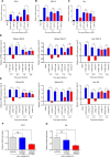
Fig 6. Effects of the PP55L amino acid substitution on the IFN response in HINAE cells and flounder.
(A–C) HINAE cells were infected with rVHSV-wild or rVHSV-P at 1 MOI, and cells were collected at the indicated time points. The expression levels of (A) IFN1, (B) ISG15, and (C) Mx in VHSV-infected HINAE cells were determined by real-time PCR. The expression levels obtained from mock-infected cells were set to 1. The results are presented as the mean ± SD of three independent experiments. *, p<0.05; **, p<0.01; ***, p<0.001; ns, not significant. (D and E) Olive flounder (43.9 ± 7.43g) were intraperitoneally injected with 2.3 × 10⁵ PFU/fish of rVHSV-wild or rVHSV-P at 13°C. At the indicated time points, kidney, spleen, and liver tissues were collected from anesthetized fish, and the expression levels of (D) ISG15 and (E) Mx in the tissues were determined by real-time PCR. The expression levels obtained from mock-infected fish were set to 1. The results are presented as the mean ± SD of three independent experiments. *, p<0.05; **, p<0.01; ***, p<0.001. ns, not significant. (F and G) HINAE cells were transfected with pcDNA6-P-wild or pcDNA6-P(P55L). Empty pcDNA6 vector was used as the control. Cells were treated with 5 μg/ml of blasticidin for 2 weeks to enrich the plasmid-bearing cells. HINAE cells transfected with pcDNA6-P-wild, pcDNA6-P(P55L), or empty pcDNA6 vector as in Fig 5 were infected with ADC-VHS2015-5 at 1 MOI. At 6 h post-infection, cells were collected and the expression levels of (F) IFN1 and (G) Mx were determined by real-time PCR. The expression levels obtained from mock-infected cells were set to 1. The results are presented as the mean ± SD of three independent experiments. *, p<0.05; ns, not significant.
Fig 7
Fig 7. Analysis of RNA-seq data from mock-, rVHSV-wild- and rVHSV-P-infected HINAE cells at 6 h post-infection.
(A) Unigenes down-regulated in rVHSV-P-infected HINAE cells compared to rVHSV-wild-infected HINAE cells were annotated to the 12 functional GO terms in the Biological Process category. (B) Fold changes in the expression of IFN response-related genes. The graph represents changes in the IFN response-related genes in rVHSV-wild- and rVHSV-P-infected cells compared to mock-infected cells. (C) HINAE cells were infected with rVHSV-wild or rVHSV-P at 1 MOI. At 6 h post-infection, cells were collected and the expression levels of IRF1A, IRF4A, IRF8, and IRF10 were determined by real-time PCR. The expression levels obtained from mock-infected cells were set to 1. The results are presented as the mean ± SD of three independent experiments. *, p<0.05; **, p<0.01; ***, p<0.001.

Similar articles

Cited by

  • Applying genetic technologies to combat infectious diseases in aquaculture.
    Robinson NA, Robledo D, Sveen L, Daniels RR, Krasnov A, Coates A, Jin YH, Barrett LT, Lillehammer M, Kettunen AH, Phillips BL, Dempster T, Doeschl-Wilson A, Samsing F, Difford G, Salisbury S, Gjerde B, Haugen JE, Burgerhout E, Dagnachew BS, Kurian D, Fast MD, Rye M, Salazar M, Bron JE, Monaghan SJ, Jacq C, Birkett M, Browman HI, Skiftesvik AB, Fields DM, Selander E, Bui S, Sonesson A, Skugor S, Østbye TK, Houston RD. Robinson NA, et al. Rev Aquac. 2023 Mar;15(2):491-535. doi: 10.1111/raq.12733. Epub 2022 Sep 5. Rev Aquac. 2023. PMID: 38504717 Free PMC article. Review.
  • Utilizing shRNA-expressing lentivectors for viral hemorrhagic septicemia virus suppression via NV gene targeting.
    Pourhoseini Dehkordi N, Saffar B, Mokhtari A, Asadi Samani L, Amini A. Pourhoseini Dehkordi N, et al. Front Vet Sci. 2025 Apr 4;12:1508470. doi: 10.3389/fvets.2025.1508470. eCollection 2025. Front Vet Sci. 2025. PMID: 40256606 Free PMC article.

References

    1. Walker PJ, Benmansour A, Calisher CH, Dietzgen R, Fang RX, Jackson AO, et al. Family Rhabdoviridae In: van Regenmortel MHV, Fauquet CM, Bishop DHL, Carstens EB, et al. editors. The seventh report of the international committee for taxonomy of viruses. San Diego: Academic Press; 2000:563–583.
    1. Schütze H, Mundt E, Mettenleiter TC. Complete genomic sequence of viral haemorrhagic septicemia virus, a fish rhabdovirus. Virus Genes. 1999;19:59–65. 10.1023/a:1008140707132 - DOI - PubMed
    1. Snow M, Cunningham CO, Melvin WT, Kurath G. Analysis of the nucleoprotein gene identifies distinct lineages of viral haemorrhagic septicaemia virus within the European marine environment. Virus Res. 1999;63:35–44. 10.1016/s0168-1702(99)00056-8 - DOI - PubMed
    1. Einer-Jensen K, Ahrens P, Forsberg R, Lorenzen N. Evolution of the fish rhabdovirus viral haemorrhagic septicaemia virus. J Gen Virol. 2004;85:1167–1179. 10.1099/vir.0.79820-0 - DOI - PubMed
    1. Lumsden JS, Morrison B, Yason C, Russell S, Young K, Yazdanpanah A, et al. Mortality event in freshwater drum Aplodinotus grunniens from Lake Ontario, Canada, associated with viral haemorrhagic septicemia virus, type IV. Dis Aquat Org. 2007;76:99–111. 10.3354/dao076099 - DOI - PubMed

Publication types

MeSH terms

LinkOut - more resources