. 2021 Jan 19;17(1):e1009191.
doi: 10.1371/journal.ppat.1009191. eCollection 2021 Jan.

Immunological design of commensal communities to treat intestinal infection and inflammation

Affiliations

Immunological design of commensal communities to treat intestinal infection and inflammation

Rebecca L Brown et al. PLoS Pathog. .

Abstract

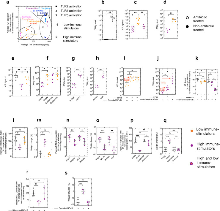
The immunological impact of individual commensal species within the microbiota is poorly understood limiting the use of commensals to treat disease. Here, we systematically profile the immunological fingerprint of commensals from the major phyla in the human intestine (Actinobacteria, Bacteroidetes, Firmicutes and Proteobacteria) to reveal taxonomic patterns in immune activation and use this information to rationally design commensal communities to enhance antibacterial defenses and combat intestinal inflammation. We reveal that Bacteroidetes and Firmicutes have distinct effects on intestinal immunity by differentially inducing primary and secondary response genes. Within these phyla, the immunostimulatory capacity of commensals from the Bacteroidia class (Bacteroidetes phyla) reflects their robustness of TLR4 activation and Bacteroidia communities rely solely on this receptor for their effects on intestinal immunity. By contrast, within the Clostridia class (Firmicutes phyla) it reflects the degree of TLR2 and TLR4 activation, and communities of Clostridia signal via both of these receptors to exert their effects on intestinal immunity. By analyzing the receptors, intracellular signaling components and transcription factors that are engaged by different commensal species, we identify canonical NF-κB signaling as a critical rheostat which grades the degree of immune stimulation commensals elicit. Guided by this immunological analysis, we constructed a cross-phylum consortium of commensals (Bacteroides uniformis, Bacteroides ovatus, Peptostreptococcus anaerobius and Clostridium histolyticum) which enhances innate TLR, IL6 and macrophages-dependent defenses against intestinal colonization by vancomycin resistant Enterococci, and fortifies mucosal barrier function during pathological intestinal inflammation through the same pathway. Critically, the setpoint of intestinal immunity established by this consortium is calibrated by canonical NF-κB signaling. Thus, by profiling the immunological impact of major human commensal species our work paves the way for rational microbiota reengineering to protect against antibiotic resistant infections and to treat intestinal inflammation.


Conflict of interest statement

The authors have declared that no competing interests exist.

Figures

Fig 1
Fig 1. Identification of commensal microbes that are robust activators of innate immunity.
(a) Cladogram of intestinal commensal species used in this study. Heat map displaying fold induction in macrophage TNF and IL6 production relative to unstimulated cells (mean values for each species are shown, n = 6–15 biological repeats, at MOI of 1:10). (b) Absolute levels of TNF produced by each commensal species ranked from low to high stimulators within each phyla. (c) Comparison of average induction of TNF and IL6 by members of the Bacteroidetes and Firmicute phyla. Each data point is mean level of cytokine production for a single commensal species, horizontal lines indicate median values. (d) Intestinal IL6 levels 24 hours post oral inoculation with 1×10⁸ CFU indicated commensal species. Each point represents a single mouse and horizontal lines indicate median values (HIGH STIMULATORS: Actinobacteria: Bifidobacterium adolescentis; Bacteroidetes: Bacteroides caccae; Firmicutes: Eubacterium infirmum; Proteobacteria: Acinetobacter radioresistens) (LOW STIMULATORS: Actinobacteria: Bifidobacterium breve; Bacteroidetes: Bacteroides cellulosilyticus; Firmicutes: Enterococcus raffinosus; Proteobacteria: Citrobacter koseri). All mice were antibiotic treated unless indicated otherwise prior to commensal administration. (e,f,g) Intestinal cytokine levels 4 (e) and 24 (f,g) hours post oral inoculation with 1×10⁸ CFU of indicated commensal consortium (BACTEROIDETES consortium: Bacteroides caccae, Bacteroides cellulosilyticus, Bacteroides dorei, Bacteroides eggerthii, Bacteroides finegoldii, Bacteroides ovatus, Bacteroides thetaiotaomicron, Bacteroides uniformis, Parabacteroides johnsonii; FIRMICUTES consortium: Clostridium histolyticum, Clostridium orbiscindens, Enterococcus faecalis, Enterococcus raffinosus, Eubacterium limosum, Faecalibacterium prausnitzii, Lactobacillus reuteri, Lactobacillus crispatus, Paenibacillus barengoltzii, Peptostreptococcus anaerobius). For (g) indicated mice were treated with clodronate liposomes or empty liposomes 2 days prior and concomitant with consortia inoculation. All mice were antibiotic treated unless indicated otherwise prior to commensal administration. Horizontal lines indicate mean values (d-g) and statistical comparisons were by Mann Whitney test *p < 0.05, **p < 0.01, and NS, not significant.
Fig 2
Fig 2. Patterns of commensal immunostimulation within the Bacteroidetes and Firmicutes phyla reflect and rely upon the activation of different TLRs.
(a) TLR-dependent SEAP production by HEK293 cells 24 hours post-stimulation with indicated commensal species (at MOI of 1:10). Corresponding frequency distribution of TLR-dependent SEAP production for all commensals in our panel. (b) Comparison of TLR activation by members of the Bacteroidetes and Firmicute phyla. Each data point is mean fold increase in TLR-dependent SEAP activity elicited by a single commensal species, horizontal lines indicate median values. (c) Correlation between overall immunostimulatory capacity of commensals in indicated phyla (measured by macrophage IL6 production (using data from Fig 1A)) and TLR-dependent SEAP activation. Correlations were determined using the Pearson’s test. (d,e) Intestinal cytokine levels 24 hours post oral inoculation with 1×10⁸ CFU of indicated commensal consortium ((d) Clostridia consortium: Eubacterium infirnum, Clostridium histolyticum, Clostridium orbiscindens, Clostridium symbiosum, Eubacterium limosum, Faecalibacterium prausnitzii, Peptostreptococcus anaerobius. (e) Bacteroidia consortium: Bacteroides caccae, Bacteroides cellulosilyticus, Bacteroides dorei, Bacteroides eggerthii, Bacteroides finegoldii, Bacteroides ovatus, Bacteroides thetaiotaomicron, Bacteroides uniformis). Indicated groups of mice were treated with TLR neutralizing antibodies, or isotype control, (100 μg/mouse) concomitant with consortia inoculation. All mice were antibiotic treated unless indicated otherwise prior to commensal administration. Each point represents a single mouse and horizontal lines indicate mean values. (b) statistical comparisons were by Mann Whitney test, (e) statistical comparisons were by Kruskal–Wallis test with Dunn’s multiple comparison test. *p < 0.05, **p < 0.01, and NS, not significant.
Fig 3
Fig 3. Canonical NF-κB is the ultimate determinant of the immunostimulatory power of commensals.
(a) Schematic of important intracellular signaling components downstream of TLRs, components inhibited in this study underlined in red. (b) Heat map displaying percentage inhibition in IL6 production after pharmacological inhibition of IRAK4 (100 nM PF06650833), P38 (100 μM SB203580), JNK (10 μM SP600125), ERK (10 μM U0126), Syk (20 μM Piceatannol), PKC (10 nM GO6983), canonical NF-κB inhibitor IKKβ (10 μM BOT-64), non-canonical NF-κB inhibitor IKKε/TBK1 (100 μM Amlexanox) and AP-1 (50 μM SR11302). Data are relative to macrophages treated with vehicle control, with mean values for percentage inhibition for each species are shown (n = 4–10 biological repeats, at MOI of 1:10). Corresponding frequency distribution of cytokine inhibition for all commensals in our panel. (c) Correlation between overall immunostimulatory capacity of all commensals in our panel (measured by macrophage IL6 production) and their dependence on the indicated signaling component (measured by cognate reduction in cytokine production incurred after inhibition of each of the intracellular signaling components). (d) Correlation between overall immunostimulatory capacity of all commensals in our panel (measured by macrophage IL6 production) and their dependence on canonical NF-κB signaling (measured by cognate reduction in cytokine production incurred after inhibition of IKKβ). Correlations were determined by Spearman’s test with False Discovery Rate correction for multiple comparisons, adjusted p-values stated with p < 0.05 considered significant.
Fig 4
Fig 4. Rational design of commensal consortia to treat intestinal infection and intestinal inflammation.
(a) Scatter plot of TLR activation (measured by TLR2-, TLR4- and TLR5-dependent SEAP activation) versus overall immunostimulatory capacity (measured by macrophage cytokine production) of indicated commensals from the Bacteroidetes and Firmicutes phyla. Commensals were grouped together into the “low immune-stimulators” (defined as <500 pg/mL TNF and <50-fold activation of all TLRs) and “high immune-stimulators” (defined as >500 pg/mL TNF and >50-fold activation of at least two TLRs). (b) VRE burden in the faeces of WT adult mice two days post-oral inoculation. Each point represents a single mouse and horizontal lines indicate median values. (c) VRE burden in the faeces of WT adult mice two days post-oral inoculation. Animals were orally inoculated with 1×10⁸ CFU of the “low immune-stimulating” commensal consortium, 1×10⁸ CFU of the “high immune-stimulating” commensal consortium, or 1×10⁸ CFU of the “low immune-stimulating” and “high immune-stimulating” commensal consortia combined, two days prior to VRE inoculation. (d) VRE burden in the faeces of WT adult mice four days post-oral inoculation. Animals were orally inoculated with 1×10⁸ CFU of the “low immune-stimulating” or “high immune-stimulating” commensal consortia two days post-VRE inoculation. (e) VRE burden in the faeces of adult SCID mice two days post-oral inoculation. Indicated animals were orally inoculated with 1×10⁸ CFU of the “low immune-stimulating” or “high immune-stimulating” commensal consortia two days prior to VRE inoculation. (f) VRE burden in the faeces of WT adult mice two days post-oral inoculation. Animals were orally inoculated with 1×10⁸ CFU of the “low immune-stimulating” or “high immune-stimulating” commensal consortia two days prior to VRE inoculation. Indicated mice were treated with clodronate liposomes or empty liposomes 2 days prior and concomitant with consortia inoculation. (g) VRE burden in the faeces of WT adult mice two days post-oral inoculation. Animals were orally inoculated with 1×10⁸ CFU of the “high immune-stimulating” commensal consortia two days prior to VRE inoculation. Concomitant with consortium inoculation mice were administered either isotype control or a cocktail of TLR neutralizing antibodies (αTLR2, αTLR4 and αTLR5, (100 μg/mouse)) via intraperitoneal injection. (h) VRE burden in the faeces of adult orally inoculated with the “high immune-stimulating” commensal consortia two days post-oral inoculation. Commensal consortia were administered two days prior to VRE inoculation. Concomitant with consortium inoculation mice were administered either isotype control or an IL6 neutralizing antibody via intraperitoneal injection (i,j) VRE burden in the faeces of WT adult mice orally inoculated with the “high immune-stimulating” commensal consortia two days post-oral inoculation with VRE. Commensal consortia were administered two days prior to VRE inoculation. For pharmacological inhibition of canonical NF-κB, mice were administered BOT-64 (30 mg/kg/dose) via intraperitoneal injection concomitant with consortia inoculation. For pharmacological inhibition of P38 mice were administered SB203580 (2 mg/kg/dose) via intraperitoneal injection concomitant with consortia inoculation. (k) Intestinal cytokine levels 24 hours post oral inoculation with 1×10⁸ CFU of indicated commensal consortium. Each point represents a single mouse and horizontal lines indicate median values. (l) Plasma FITC-dextran levels in mice with DSS-induced intestinal inflammation. For the induction of intestinal inflammation, antibiotic treated mice received 2% (w/v) DSS in drinking water for seven days. Indicated groups of mice were orally administered commensal consortia on day 2, day 4 and day 6 of DSS treatment. One day after cessation of DSS treatment, mice were orally administered 200 μL of FITC-dextran 4000 (80 mg/mL) and FITC-dextran 4000 levels in blood were measured after four hours. (m) Weight change in DSS-treated mice six days after start of DSS treatment. (n) Plasma FITC-dextran levels in mice with DSS-induced intestinal inflammation. For the induction of intestinal inflammation, antibiotic treated mice received 2% (w/v) DSS in drinking water for seven days. Indicated groups of mice were orally administered commensal consortia on day 2, day 4 and day 6 of DSS treatment. One day after cessation of DSS treatment, mice were orally administered 200 μL of FITC-dextran 4000 (80 mg/mL) and FITC-dextran 4000 levels in blood were measured after four hours. In indicated groups, concomitant with consortium inoculation mice were administered either isotype control or a cocktail of TLR neutralizing antibodies (αTLR2, αTLR4 and αTLR5, (100 μg/mouse)) or IL6 neutralizing antibody (100 μg/mouse), or isotype control via intraperitoneal injection. (o) Weight change in DSS-treated mice six days after start of DSS treatment. In indicated groups, concomitant with consortium inoculation mice were administered either isotype control or a cocktail if TLR neutralizing antibodies (αTLR2, αTLR4 and αTLR5, (100 μg/mouse)) or IL6 neutralizing antibody (100 μg/mouse), or isotype control via intraperitoneal injection. (p) Plasma FITC-dextran levels in mice with DSS-induced intestinal inflammation. For the induction of intestinal inflammation, antibiotic treated mice received 2% (w/v) DSS in drinking water for seven days. Indicated groups of mice were orally administered commensal consortia on day 2, day 4 and day 6 of DSS treatment. One day after cessation of DSS treatment, mice were orally administered 200 μL of FITC-dextran 4000 (80 mg/mL) and FITC-dextran 4000 levels in blood were measured after four hours. In indicated groups, concomitant with consortium inoculation, mice were treated with clodronate liposomes or empty liposomes concomitant with consortia inoculation. (q) Weight change in DSS-treated mice six days after start of DSS treatment. In indicated groups, concomitant with consortium inoculation, mice were treated with clodronate liposomes or empty liposomes concomitant with consortia inoculation. Each point represents a single mouse and horizontal lines indicate mean values. (r) Plasma FITC-dextran levels in mice with DSS-induced intestinal inflammation. For the induction of intestinal inflammation, antibiotic treated mice received 2% (w/v) DSS in drinking water for seven days. Indicated groups of mice were orally administered commensal consortia on day 2, day 4 and day 6 of DSS treatment. One day after cessation of DSS treatment, mice were orally administered 200 μL of FITC-dextran 4000 (80 mg/mL) and FITC-dextran 4000 levels in blood were measured after four hours. In indicated groups, for pharmacological inhibition of canonical NF-κB, mice were administered BOT-64 (30 mg/kg/dose) via intraperitoneal injection concomitant with consortia inoculation concomitant with consortium inoculation. (s) Weight change in DSS-treated mice six days after start of DSS treatment. In indicated groups, for pharmacological inhibition of canonical NF-κB, mice were administered BOT-64 (30 mg/kg/dose) via intraperitoneal injection concomitant with consortia inoculation concomitant with consortium inoculation. (b-h) Statistical comparisons were by Mann Whitney test, (i-s) statistical comparisons were by Kruskal–Wallis test with Dunn’s multiple comparison test. *p < 0.05, **p < 0.01, and NS, not significant.

Similar articles

Cited by

References

    1. Brown RL, Clarke TB. The regulation of host defences to infection by the microbiota. Immunology. 2017;150(1):1–6. 10.1111/imm.12634 - DOI - PMC - PubMed
    1. Honda K, Littman DR. The microbiome in infectious disease and inflammation. Annu Rev Immunol. 2012;30:759–95. 10.1146/annurev-immunol-020711-074937 - DOI - PMC - PubMed
    1. Kawai T, Akira S. The role of pattern-recognition receptors in innate immunity: update on Toll-like receptors. Nat Immunol. 2010;11(5):373–84. 10.1038/ni.1863 - DOI - PubMed
    1. Baumler AJ, Sperandio V. Interactions between the microbiota and pathogenic bacteria in the gut. Nature. 2016;535(7610):85–93. 10.1038/nature18849 - DOI - PMC - PubMed
    1. Cho I, Blaser MJ. The human microbiome: at the interface of health and disease. Nat Rev Genet. 2012;13(4):260–70. 10.1038/nrg3182 - DOI - PMC - PubMed

Publication types

MeSH terms

LinkOut - more resources