. 2021 Jan 19;17(1):e1009212.
doi: 10.1371/journal.ppat.1009212. eCollection 2021 Jan.

Hydroxychloroquine-mediated inhibition of SARS-CoV-2 entry is attenuated by TMPRSS2

Affiliations

Hydroxychloroquine-mediated inhibition of SARS-CoV-2 entry is attenuated by TMPRSS2

Tianling Ou et al. PLoS Pathog. .

Abstract

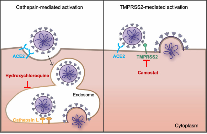
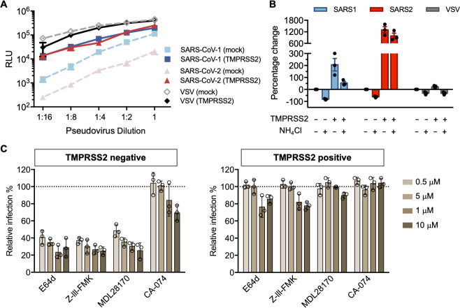
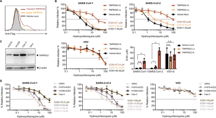
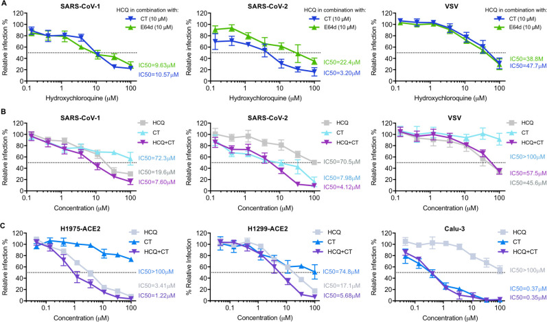
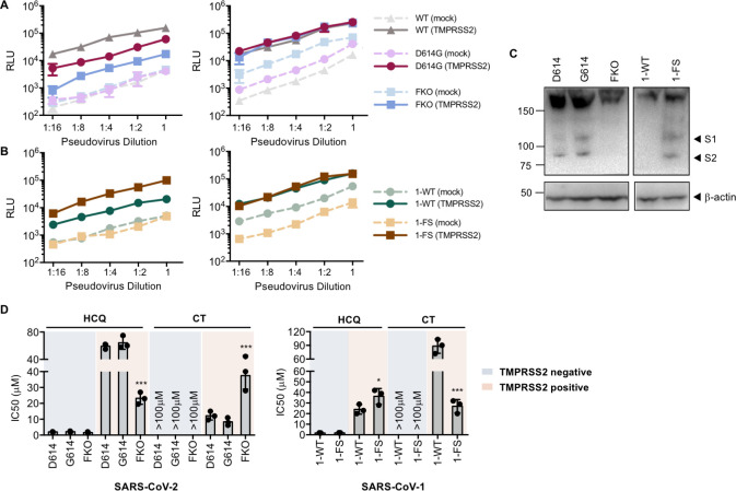
Hydroxychloroquine, used to treat malaria and some autoimmune disorders, potently inhibits viral infection of SARS coronavirus (SARS-CoV-1) and SARS-CoV-2 in cell-culture studies. However, human clinical trials of hydroxychloroquine failed to establish its usefulness as treatment for COVID-19. This compound is known to interfere with endosomal acidification necessary to the proteolytic activity of cathepsins. Following receptor binding and endocytosis, cathepsin L can cleave the SARS-CoV-1 and SARS-CoV-2 spike (S) proteins, thereby activating membrane fusion for cell entry. The plasma membrane-associated protease TMPRSS2 can similarly cleave these S proteins and activate viral entry at the cell surface. Here we show that the SARS-CoV-2 entry process is more dependent than that of SARS-CoV-1 on TMPRSS2 expression. This difference can be reversed when the furin-cleavage site of the SARS-CoV-2 S protein is ablated or when it is introduced into the SARS-CoV-1 S protein. We also show that hydroxychloroquine efficiently blocks viral entry mediated by cathepsin L, but not by TMPRSS2, and that a combination of hydroxychloroquine and a clinically-tested TMPRSS2 inhibitor prevents SARS-CoV-2 infection more potently than either drug alone. These studies identify functional differences between SARS-CoV-1 and -2 entry processes, and provide a mechanistic explanation for the limited in vivo utility of hydroxychloroquine as a treatment for COVID-19.


Conflict of interest statement

The authors have declared that no competing interests exist.

Figures

Fig 1
Fig 1. SARS-CoV-1 and SARS-CoV-2 fusion can be activated by either or both of two pathways.
The coronaviruses bind the cellular receptor ACE2 and must be activated by proteolysis with either a surface-expressed protease like TMPRSS2 or by cathepsin L in the endosome. Only cathepsin L-mediated proteolysis requires endosomal acidification. Camostat mesylate inhibits TMPRSS2 activity, whereas hydroxychloroquine, like ammonium chloride, inhibits endosomal acidification.
Fig 2
Fig 2. SARS-CoV-2 is resistant to endosomal protease inhibitors in cells over-expressing TMPRSS2.
(A) 293T-ACE2 cells were transfected with a vector control plasmid (TMPRSS2-negative), or a TMPRSS2 expression plasmid, 24 hours prior to infection. Retroviruses pseudotyped with SARS-CoV-1 (SARS1), SARS-CoV-2 (SARS2) S, and VSV G proteins were used for infection at titers yielding equivalent infection in the presence of TMPRSS2. Pseudovirus was serially diluted by 2-fold starting from stock (RLU≈250,000 for TMPRSS2-expressing cells). Cells were inoculated with diluted pseudovirus, and viral entry was determined by the luciferase activity in cell lysates within 48 hours post infection. Shown is a representative plot from three experiments. Each point indicates the mean (±SD) of duplicate samples. (B) Cells were treated by 50 mM of ammonium chloride before infection. Infection of TMPRSS2-negative cells without treatment was used for normalization. (C) A panel of cathepsin inhibitors was tested: E64d and Z-III-FMK inhibit both cathepsin B and cathepsin L; MDL28170 specifically inhibits cathepsin L, and CA-074 inhibits cathepsin B. Cells were treated with the indicated concentrations of protease inhibitors or DMSO for 2 hours, then inoculated with retrovirus harboring SARS-CoV-2 spike proteins. Luciferase activity was measured at 48 hours post inoculation. Relative infection (%) was calculated from infection of DMSO-treated cells. Each point in (B) and (C) represents the mean of triplicate samples from one experiment. Bars indicate the average of three independent experiments and error bars indicate SD.
Fig 3
Fig 3. Antiviral effect of hydroxychloroquine is dependent on TMPRSS2 expression.
(A) Cell-surface staining was performed by detecting the flag tag at the C-terminal of TMPRSS2 to validate the overexpression of TMPRSS2. A stable cell line of 293T-ACE2 cells was generated to express TMPRSS2 (orange line). 293T-ACE2 cells were transiently transfected with a vector control (black line), or TMPRSS2 (red line). The 293T/ACE2/TMPRSS2 stable cell line had much lower expression of TMPRSS2 compared to 293T-ACE2 transiently transfected with TMPRSS2 plasmids, and thus were referred as TMPRSS2 Lo and TMPRSS2 Hi, respectively. Shown is a representation of flow cytometry data from two independent experiments. (B) 293-ACE2 cells with different levels of TMPRSS2 were treated with hydroxychloroquine or DMSO before virus inoculation. The results are presented as a percentage of infection of DMSO-treated cells (≈ 250,000 RLU for all three pseudotypes from TMPRSS-expressing cells.) Shown are representative plots of the mean value (±SD) of triplicate samples from the viral entry inhibition assay. The bar graph is a summary of IC50 from three independent experiments. Each point represents the IC50 calculated from one experiment. Unpaired Student’s t-test was used to assess the statistical significance of the difference between IC50 on mock transfected cells (TMPRSS2-negative) and TMPRSS2-positive cells. (**: P < 0.01. *: P < 0.05. n.s.: P > 0.05.) (C) Shown is a representative western blot (of two independent experiments performed) reflecting expression level of TMPRSS2 in different cell lines. H1299, H1975 and Calu-3: human non-small cell lung cancer cells; Vero: kidney epithelial cells. β-Actin served as loading controls. (D) Lentiviruses pseudotyped with SARS-CoV-1, SARS-CoV-2 S, and VSV G proteins were used for infection. Stable cells lines of H1299 and H1975 over-expressing ACE2 were generated to allow efficient infection. Drug treatment, virus inoculation, and luciferase measurement were the same with the procedures in (B). Shown are the mean value (±SD) of triplicate samples from three independent experiments.
Fig 4
Fig 4. Suppression of TMPRSS2 restores the antiviral efficiency of hydroxychloroquine.
(A) Hydroxychloroquine (HCQ) and camostat (CT) were tested on 293T-ACE2 cells transiently expressing TMPRSS2 prior to infection. HCQ was serially diluted with complete media containing 10 μM of camostat (CT, blue solid line), or 10 μM of E64d (cyan solid line), respectively. The antiviral efficiency of hydroxychloroquine in combination of each inhibitor was compared. (B-C) The antiviral efficiency of HCQ and CT alone, and their combination, were tested on (B) 293T-ACE2 cells and (C) other human lung cell lines (H1975-ACE2, H1299-ACE2, and Calu-3). Cells were challenged with retroviruses pseudotyped with SARS-CoV-2, SARS-CoV-1 S proteins, and VSV-G in (B), and with lentivirus pseudotyped with SARS-CoV-2 S proteins in (C), after drug or DMSO treatment. Luciferase activity was measured at 48 hours post inoculation. The average of three independent experiments conducted with triplicates is shown in (A–C). Error bars indicate SD. Relative infection (%) was calculated from infection of DMSO-treated cells.
Fig 5
Fig 5. Furin cleavage in the virion producer cell correlates with TMPRSS2 dependence.
(A-B) The infectivity of SARS-CoV-1 and SARS-CoV-2 and their mutants on 293-ACE2 cells was compared with or without overexpression of TMPRSS2. The infection assay was performed as described in Fig 2A. Retroviruses were pseudotyped with S proteins of SARS-CoV-2 wildtype (WT), D614G S-protein variant, SARS-CoV-2 with furin-site knockout (FKO), and (B) SARS-CoV-1 WT (1-WT), and SARS-CoV-1 with the furin site derived from SARS-CoV-2 (1-FS). Pseudovirus titers were adjusted so that they were equivalent in the absence (left panel) or presence (right panel) of TMPRSS2. Shown is a representative plot of the mean (±SD) of duplicate samples from at least two independent experiments. (C) Western blot analysis of S protein cleavage of SARS-CoV-2-S WT (D614) and variants (G614 and FKO), and SARS-CoV-1 WT and its variant with furin site addition. Antibody detected the flag tags at the N- and C-termini of S proteins. β-Actin served as loading controls. Black arrow heads indicate bands corresponding to the S1 and S2 subunits from cleavage of S proteins. Shown are representative blots from two experiments. (D)The effect of TMPRSS2 on the antiviral efficiency of hydroxychloroquine (HCQ) and camostat was compared. Retrovirus pseudotyped against S proteins described in (C) after drug or DMSO treatment. Luciferase activity was measured at 48 hours post inoculation. Relative infection (%) was calculated from infection of DMSO-treated cells. Statistical significance between wildtypes and mutants was tested by two-way ANOVA with Dunnett’s posttest. (***: P < 0.001. *: P < 0.05).

Similar articles

Cited by

References

    1. Spinelli A, Pellino G. COVID-19 pandemic: perspectives on an unfolding crisis. The British Journal of Surgery. 2020. - PMC - PubMed
    1. Sanders JM, Monogue ML, Jodlowski TZ, Cutrell JB. Pharmacologic treatments for coronavirus disease 2019 (COVID-19): a review. Jama. 2020;323(18):1824–36. 10.1001/jama.2020.6019 - DOI - PubMed
    1. Geleris J, Sun Y, Platt J, Zucker J, Baldwin M, Hripcsak G, et al. Observational study of hydroxychloroquine in hospitalized patients with Covid-19. New England Journal of Medicine. 2020. 10.1056/NEJMoa2012410 - DOI - PMC - PubMed
    1. Chen J, Liu D, Liu L, Liu P, Xu Q, Xia L, et al. A pilot study of hydroxychloroquine in treatment of patients with moderate COVID-19. Zhejiang da xue xue bao Yi xue ban = Journal of Zhejiang University Medical sciences. 2020;49(2):215 10.3785/j.issn.1008-9292.2020.03.03 - DOI - PMC - PubMed
    1. Chen Z, Hu J, Zhang Z, Jiang S, Han S, Yan D, et al. Efficacy of hydroxychloroquine in patients with COVID-19: results of a randomized clinical trial. MedRxiv. 2020. 10.1001/jama.2020.22240 - DOI - PMC - PubMed

Publication types

MeSH terms