. 2021 Mar:333:108543.
doi: 10.1016/j.mbs.2021.108543. Epub 2021 Jan 16.

A simple model of immune and muscle cell crosstalk during muscle regeneration

Affiliations

A simple model of immune and muscle cell crosstalk during muscle regeneration

Hristo V Kojouharov et al. Math Biosci. 2021 Mar.

Abstract

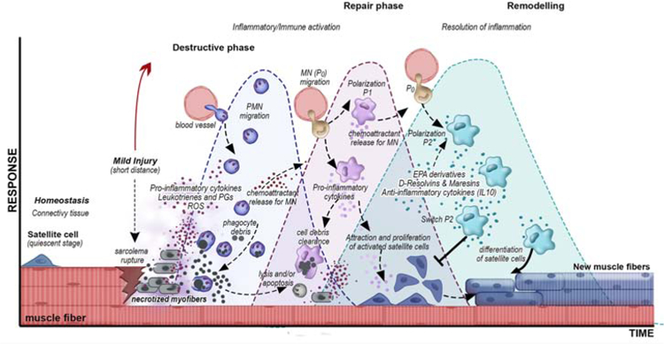
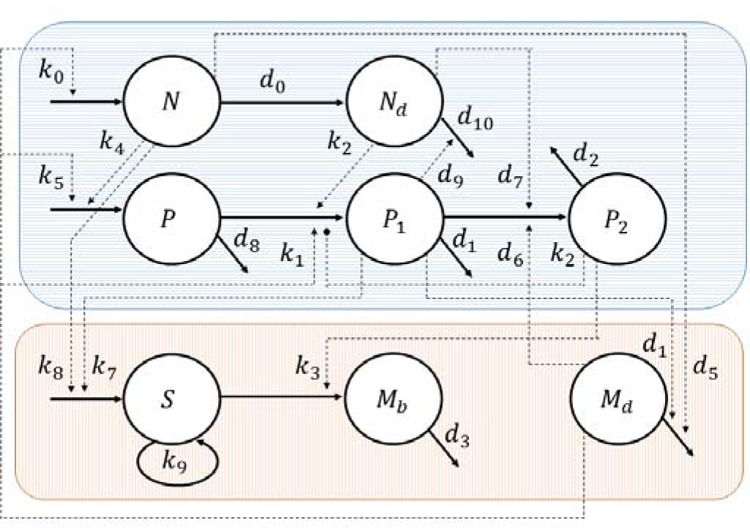
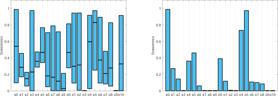
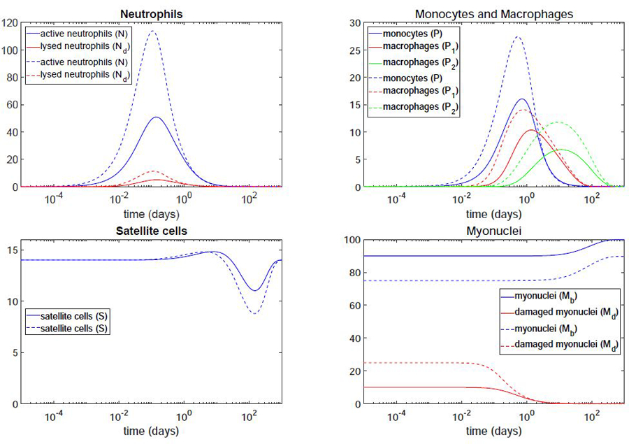
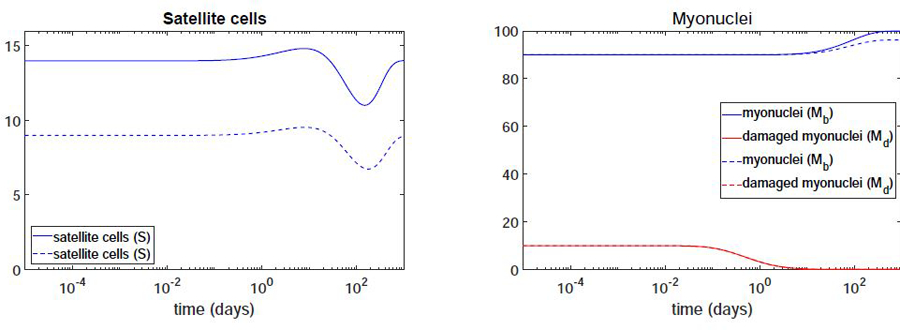
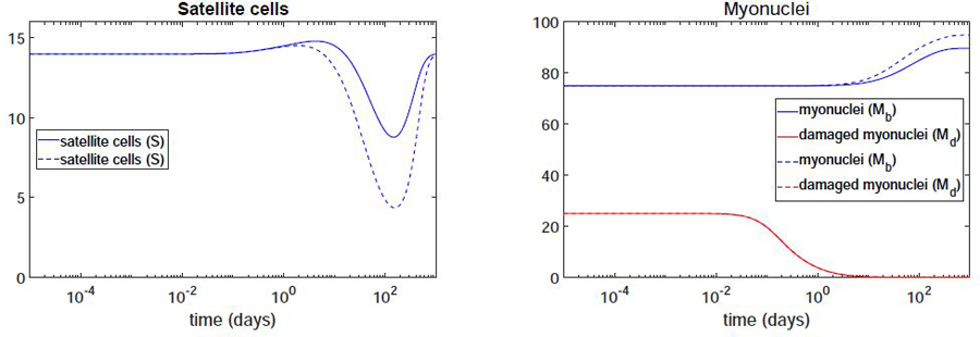
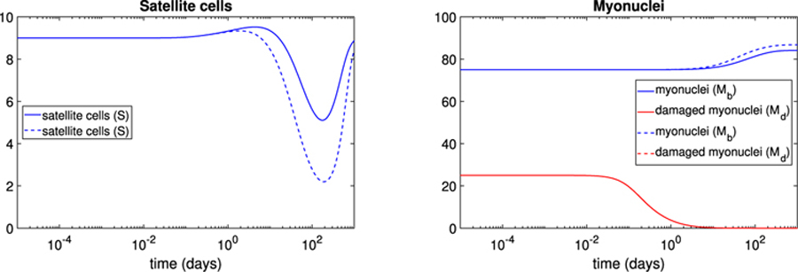
Muscle injury during aging predisposes skeletal muscles to increased damage due to reduced regenerative capacity. Some of the common causes of muscle injury are strains, while other causes are more complex muscle myopathies and other illnesses, and even excessive exercise can lead to muscle damage. We develop a new mathematical model based on ordinary differential equations of muscle regeneration. It includes the interactions between the immune system, healthy and damaged myonuclei as well as satellite cells. Our new mathematical model expands beyond previous ones by accounting for 21 specific parameters, including those parameters that deal with the interactions between the damaged and dead myonuclei, the immune system, and the satellite cells. An important assumption of our model is the replacement of only damaged parts of the muscle fibers and the dead myonuclei. We conduce systematic sensitivity analysis to determine which parameters have larger effects on the model and therefore are more influential for the muscle regeneration process. We propose additional validation for these parameters. We further demonstrate that these simulations are species-, muscle-, and age-dependent. In addition, the knowledge of these parameters and their interactions, may suggest targeting or selecting these interactions for treatments that accelerate the muscle regeneration process.

Keywords: Mathematical modeling; Muscle; Myotubes; Regeneration.


Conflict of interest statement

Declaration of Competing Interest The authors declare that they have no known competing financial interests or personal relationships that could have appeared to influence the work reported in this paper.

Figures

Figure 1:
Figure 1:
Schematic illustration of the muscle response to injury and the phases of muscle regeneration/repair. At the lower part of the figure in dark pink color is the muscle fiber (myofiber). In this example, as indicated by the black and red arrows, a mild to moderate injury occurs, which leads to muscle cell damage resulting in sarcolemma rupture, triggering the inflammatory/immune activation in the destructive phase of the response. This initial phase is highly dominated by proinflammatory molecules, such as cytokines, reactive oxygen species (ROS), and lipid signaling mediators (LMs, i.e. leukotrienes and prostaglandins [PGs]). Peripheral Mononuclear Monocytes (PMNs) are the first cells attracted to site of injury and begin phagocytosis of the muscle cell debris. As the destructive phase evolves to the repair phase, the non-polarized (denoted by P) monocytes (MN) migrate from peripheral circulation towards the site of injury and polarize into classically activated macrophages (denoted by P1) in response to LMs released from PMN. This evolution of the destructive phase to the repair phase is commonly refereed to as the the remodeling phase of muscle regeneration. P1 release contributes to the production of pro-inflammatory cytokines, attraction and proliferation of activated satellite cells, phagocytosis/clearance of residual debris from necrotic tissue and PMNs. In the late stage of the repair phase, the production of anti-inflammatory cytokines (IL-10) and LMs (EPA derivatives, D-Resolvins, and Maresins) regulate the resolution of inflammation and new muscle fibers differentiation. These molecules contribute to the switch from classically to alternatively activated macrophage polarization (denoted by P2), which ultimately inhibit the proliferation of satellite cells and induce their differentiation into new muscle fibers.
Figure 2:
Figure 2:
Flow diagram of the immune (top) and muscle (bottom) cell cross-talk during muscle regeneration.
Figure 3:
Figure 3:
Maximum, minimum and mean of the indices over all species of the indices averaged over one hundred days (left); Indices for the damaged and dead myonuclei averaged over one hundred days (right).
Figure 4:
Figure 4:
Log-Linear plots of the immune (top) and muscle (bottom) cells. Simulation results for younger adults with type I muscle, S0 = 14, under muscle damage of 10% (solid lines) and 25% (dashed lines).
Figure 5:
Figure 5:
Log-Linear plots of the muscle cells: satellite cells (left) and myonuclei (right). Simulation results for younger adults with type I muscle, S0 = 14, (solid lines) and older adults with type II muscle, S0 = 9, (dashed lines) under muscle damage of 10%.
Figure 6:
Figure 6:
Log-Linear plots of the muscle cells: satellite cells (left) and myonuclei (right). Simulation results for younger adults with type I muscle, S0 = 14, under muscle damage of 25% for different differentiation rates of satellite cells to myonuclei: k3 = 0.001 (solid lines) and k3 = 0.001 (dashed lines).
Figure 7:
Figure 7:
Log-Linear plots of the muscle cells: satellite cells (left) and myonuclei (right). Simulation results for older adults with type II muscle, S0 = 9, under muscle damage of 25% for different differentiation rates of satellite cells to myonuclei: k3 = 0.001 (solid lines) and k3 = 0.001 (dashed lines).

Similar articles

Cited by

References

    1. Karalaki M, Fili S, Philippou A, Koutsilieris M, Muscle Regeneration: Cellular and Molecular Events, In Vivo 23 (5) (2009) 779–796. - PubMed
    1. Clarkson PM, Hubal MJ, Exercise-induced muscle damage in humans, American Journal of Physical Medicine & Rehabilitation 81 (11) (2002) S52–S69. doi:10.1097/00002060-200211001-00007. - DOI - PubMed
    1. Brotto M, Abreu EL, Sarcopenia: Pharmacology of today and tomorrow, Journal of Pharmacology and Experimental Therapeutics 343 (3) (2012) 540–546. doi:10.1124/jpet.112.191759. - DOI - PMC - PubMed
    1. Choi S-J, Age-related functional changes and susceptibility to eccentric contraction-induced damage in skeletal muscle cell, Integrative Medicine Research 5 (3) (2016) 171–175, special Issue - IMPACT (Integrative Medicine: Physical Activity is a Core Tip). doi:10.1016/j.imr.2016.05.004. - DOI - PMC - PubMed
    1. Booth FW, Roberts CK, Laye MJ, Lack of Exercise Is a Major Cause of Chronic Diseases, American Cancer Society, 2012, Ch. 2, pp. 1143–1211. doi:10.1002/cphy.c110025. - DOI - PMC - PubMed

Publication types

MeSH terms

LinkOut - more resources