. 2021 May;97(3):517-523.
doi: 10.1111/php.13383. Epub 2021 Feb 5.

Exposure of Human Skin Models to KrCl Excimer Lamps: The Impact of Optical Filtering

Affiliations

Exposure of Human Skin Models to KrCl Excimer Lamps: The Impact of Optical Filtering

Manuela Buonanno et al. Photochem Photobiol. 2021 May.

Abstract

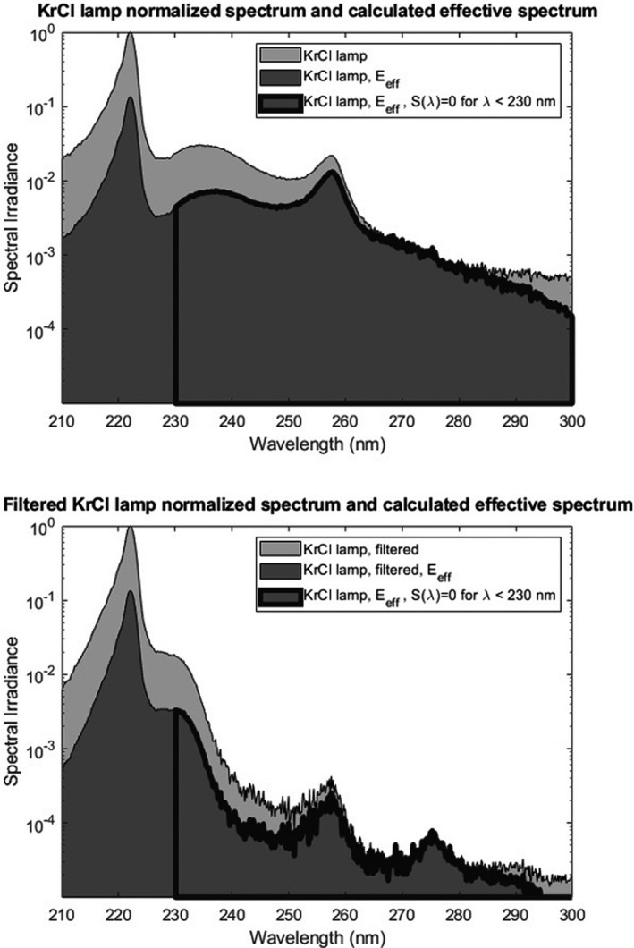
Far-UVC radiation is a promising technology that is potentially both effective at killing airborne microbes such as coronaviruses and influenza, and being minimally hazardous to the skin and eyes. Our previous studies on health risks from far-UVC have employed a krypton-chloride (KrCl) excimer lamp, emitting principally at 222 nm, supplemented with an optical filter to remove longer wavelength emissions inherent to these lamps. This study explores KrCl lamp health hazards by comparing filtered and unfiltered KrCl lamps using effective spectral irradiance calculations and experimental skin exposures. Analysis of effective irradiances showed a notable increase in allowable exposure when using a filter. Induction of DNA dimers (CPD and 6-4PP) was measured in human skin models exposed to a range of radiant exposures up to 500 mJ cm-2 . Compared to sham-exposed tissues, the unfiltered KrCl lamps induced a statistically significant increase in the yield of both DNA lesions at all the radiant exposures studied. Conversely, filtered KrCl lamps do not induce increased levels of dimers at the current daily TLV exposure limit for 222 nm (23 mJ cm-2 ). This work supports the use of filters for far-UVC KrCl excimer lamps when used to limit disease transmission in occupied locations.


Conflict of interest statement

The authors declare the following pending patent: Patent Title: “Apparatus, method and system for selectively affecting and/or killing a virus”. Applicant: The Trustees of Columbia University in the City of New York. Inventors: Gerhard Randers‐Pehrson, David Jonathan Brenner, Alan Bigelow. Application #: US20180169279A1. Aspect of manuscript covered in patent application: Spectrum filtering elements such as multilayer dielectric filters or chemical filters are used to remove unwanted wavelengths, or those wavelengths that can be outside of the preferable range of wavelengths. URL: https://patents.google.com/patent/US20180169279A1/en?oq=20200085984

Figures

Figure 1
Figure 1
Plot of the spectral irradiance of a KrCl lamp normalized to the 222 nm peak (–). Also plotted is the normalized spectral irradiance of the KrCl lamp with the addition of a custom filter that reduces the intensity outside of the 222 nm peak (+).
Figure 2
Figure 2
Normalized spectral irradiance of an unfiltered (top) and filtered (bottom) KrCl lamp along with the calculated effective spectrum. Also included on each plot are the effective spectral areas used for the hypothetical situation where the hazard function S(λ) has a value of zero for wavelengths below 230 nm.
Figure 3
Figure 3
Cyclobutane pyrimidine dimers (CPD) yields induced by filtered and unfiltered 222‐nm UV radiation in 3‐D human skin tissue models. Representative cross‐sectional images of tissue models comparing premutagenic skin lesions CPD (dark‐stained cells) in the epidermis of sham‐exposed samples (a), of samples exposed to 23, 50, 150, or 500 mJ cm−2 from filtered (b‐f) or unfiltered 222‐nm radiation (g–k). (l) Quantification of the percentage of keratinocytes showing CPD dimers. Values represent the average ± SEM of cells exhibiting dimers measured in at least six randomly selected fields of view per samples (n = 2 or 3; an average of ~ 1470 cells per sample were counted); *P < 0.001, #P < 0.05.
Figure 4
Figure 4
6‐4 pyrimidine pyrimidone dimers (6‐4PP) yields induced by filtered and unfiltered 222‐nm UV radiation in 3‐D human skin tissue models. Representative cross‐sectional images of tissue models comparing premutagenic skin lesions 6‐4PP (dark‐stained cells) in the epidermis of sham‐exposed samples (a), of samples exposed to 23, 50, 150, or 500 mJ cm−2 from filtered (b–f) or unfiltered (g–k) 222‐nm radiation. (l) Quantification of the percentage of keratinocytes showing 6‐4PP dimers. Values represent the average ± SEM of cells exhibiting dimers measured in at least six randomly selected fields of view per samples (n = 2 or 3; an average of ~ 1320 cells per sample were counted); *P ≤ 0.001, #P < 0.05.
Figure 5
Figure 5
Premutagenic DNA lesion yields induced on 3‐D human skin tissues by 222‐ or 254‐nm UV radiation at the corresponding current regulatory dose limits. Representative cross‐sectional images of samples comparing premutagenic skin lesions CPD ((a–d), dark‐stained cells), and 6‐4PP ((e‐h), dark‐stained cells) in the epidermis of sham‐exposed skin ((a) for CPD and (e) for 6‐4PP); of samples exposed 6 mJ cm−2 from 254‐nm radiation ((b) for CPD and (f) for 6‐4PP); of samples exposed to 23 or mJ cm−2 from filtered ((c) for CPD and (g) for 6‐4PP) or unfiltered 222‐nm radiation ((d) for CPD and (h) for 6‐4PP). Quantification of the percentage of keratinocytes showing CPD (i) and 6‐4PP dimers (j). Values represent the average ± SEM of cells exhibiting dimers measured in at least six randomly selected fields of view per mouse (n = 2; an average of ~ 1335 and 1170 cells per sample were counted for CPD and 6‐4PP, respectively); § P < 0.005, *P < 0.001, **P ≤ 0.0001.

References

    1. Narita, K. , Asano K., Naito K., Ohashi H., Sasaki M., Morimoto Y., Igarashi T. and Nakane A. (2020) 222‐nm UVC inactivates a wide spectrum of microbial pathogens. J Hosp Infect. 105(3), 459–467. - PubMed
    1. Wang, D. , Oppenländer T., El‐Din M. G. and Bolton J. R. (2010) Comparison of the disinfection effects of vacuum‐UV (VUV) and UV light on Bacillus subtilis spores in aqueous suspensions at 172, 222 and 254 nm. Photochem Photobiol. 86(1), 176–181. - PubMed
    1. Kang, J. W. , Kim S. S. and Kang D. H. (2018) Inactivation dynamics of 222 nm krypton‐chlorine excilamp irradiation on Gram‐positive and Gram‐negative foodborne pathogenic bacteria. Food Res Int. 109, 325–333. - PubMed
    1. Taylor, W. , Camilleri E., Craft D. L., Korza G., Granados M. R., Peterson J., Szczpaniak R., Weller S. K., Moeller R., Douki T., Mok W. W. K. and Setlow P. (2020) DNA damage kills bacterial spores and cells exposed to 222‐nanometer UV radiation. Appl. Environ Microbiol. 86(8). - PMC - PubMed
    1. Matafonova, G. G. , Batoev V. B., Astakhova S. A., Gómez M. and Christofi N. (2008) Efficiency of KrCl excilamp (222 nm) for inactivation of bacteria in suspension. Lett. Appl. Microbiol. 47(6), 508–513. - PubMed

Publication types

LinkOut - more resources