

Temporomandibular Joint Osteoarthritis: Regenerative Treatment by a Stem Cell Containing Advanced Therapy Medicinal Product (ATMP)-An In Vivo Animal Trial
- PMID: 33466246
- PMCID: PMC7795212
- DOI: 10.3390/ijms22010443
Temporomandibular Joint Osteoarthritis: Regenerative Treatment by a Stem Cell Containing Advanced Therapy Medicinal Product (ATMP)-An In Vivo Animal Trial
Abstract
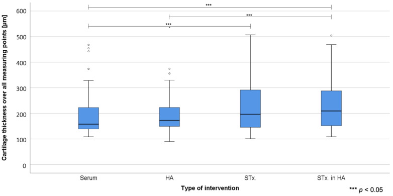
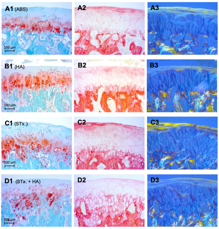
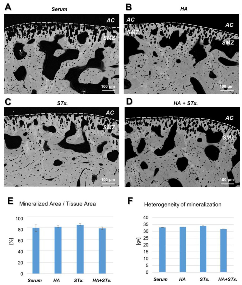
Temporomandibular joint osteoarthritis (TMJ-OA) is a chronic degenerative disease that is often characterized by progressive impairment of the temporomandibular functional unit. The aim of this randomized controlled animal trial was a comparative analysis regarding the chondroregenerative potency of intra-articular stem/stromal cell therapy. Four weeks after combined mechanical and biochemical osteoarthritis induction in 28 rabbits, therapy was initiated by a single intra-articular injection, randomized into the following groups: Group 1: AB Serum (ABS); Group 2: Hyaluronic acid (HA); Group 3: Mesenchymal stromal cells (STx.); Group 4: Mesenchymal stromal cells in hyaluronic acid (HA + STx.). After another 4 weeks, the animals were euthanized, followed by histological examination of the removed joints. The histological analysis showed a significant increase in cartilage thickness in the stromal cell treated groups (HA + STx. vs. ABS, p = 0.028; HA + ST.x vs. HA, p = 0.042; STx. vs. ABS, p = 0.036). Scanning electron microscopy detected a similar heterogeneity of mineralization and tissue porosity in the subchondral zone in all groups. The single intra-articular injection of a stem cell containing, GMP-compliant advanced therapy medicinal product for the treatment of iatrogen induced osteoarthritis of the temporomandibular joint shows a chondroregenerative effect.
Keywords: TMJD; osteoarthritis; regenerative medicine; regenerative therapy; stem cell therapy; temporomandibular joint.
Conflict of interest statement
M.K. is associated with Oxacell® AG but had no role in the design of the study, in the collection, analysis or interpretation of the data, in the writing of the manuscript or in the decision to publish the results. All other authors declare no conflict of interest.
Figures
Similar articles
-
Temporomandibular Joint Osteoarthritis: Pathogenic Mechanisms Involving the Cartilage and Subchondral Bone, and Potential Therapeutic Strategies for Joint Regeneration.Int J Mol Sci. 2022 Dec 22;24(1):171. doi: 10.3390/ijms24010171. Int J Mol Sci. 2022. PMID: 36613615 Free PMC article. Review.
-
Preliminary evaluation of histological changes found in a mechanical arthropatic temporomandibular joint (TMJ) exposed to an intra-articular Hyaluronic acid (HA) injection, in a rat model.J Craniomaxillofac Surg. 2011 Dec;39(8):610-4. doi: 10.1016/j.jcms.2010.12.001. Epub 2011 Jan 8. J Craniomaxillofac Surg. 2011. PMID: 21216612
-
Can intra-articular daidzein injection reduce oxidative damage and early osteoarthritis in a rabbit temporomandibular joint model?BMC Oral Health. 2024 Oct 8;24(1):1193. doi: 10.1186/s12903-024-04990-4. BMC Oral Health. 2024. PMID: 39379866 Free PMC article.
-
FGF18 induces chondrogenesis and anti-osteoarthritic effects in a mouse model for TMJ degeneration.PLoS One. 2025 Apr 24;20(4):e0317816. doi: 10.1371/journal.pone.0317816. eCollection 2025. PLoS One. 2025. PMID: 40273050 Free PMC article.
-
Emerging Potential of Exosomes in Regenerative Medicine for Temporomandibular Joint Osteoarthritis.Int J Mol Sci. 2020 Feb 24;21(4):1541. doi: 10.3390/ijms21041541. Int J Mol Sci. 2020. PMID: 32102392 Free PMC article. Review.
Cited by
-
Advances in Regenerative Dentistry Approaches: An Update.Int Dent J. 2024 Feb;74(1):25-34. doi: 10.1016/j.identj.2023.07.008. Epub 2023 Aug 2. Int Dent J. 2024. PMID: 37541918 Free PMC article. Review.
-
ZC3H13 Promotes NSUN4-Mediated Chondrocyte Mitochondrial Dysfunction and Pyroptosis in Temporomandibular Joint Osteoarthritis.Cartilage. 2025 May 28:19476035251339410. doi: 10.1177/19476035251339410. Online ahead of print. Cartilage. 2025. PMID: 40433805 Free PMC article.
-
Mesenchymal Stem Cell-Based Therapies for Temporomandibular Joint Repair: A Systematic Review of Preclinical Studies.Cells. 2024 Jun 6;13(11):990. doi: 10.3390/cells13110990. Cells. 2024. PMID: 38891122 Free PMC article.
-
Eosinophils-Induced Lumican Secretion by Synovial Fibroblasts Alleviates Cartilage Degradation via the TGF-β Pathway Mediated by Anxa1 Binding.Adv Sci (Weinh). 2025 Aug;12(29):e2416030. doi: 10.1002/advs.202416030. Epub 2025 Mar 24. Adv Sci (Weinh). 2025. PMID: 40125663 Free PMC article.
-
Temporomandibular Joint Osteoarthritis: Pathogenic Mechanisms Involving the Cartilage and Subchondral Bone, and Potential Therapeutic Strategies for Joint Regeneration.Int J Mol Sci. 2022 Dec 22;24(1):171. doi: 10.3390/ijms24010171. Int J Mol Sci. 2022. PMID: 36613615 Free PMC article. Review.
References
-
- Gopal K., Shankar R., Vardhan H. Prevalence of temporo‑mandibular joint disorders in symptomatic and asymptomatic patients: A cross‑sectional study. Int. J. Adv. Sci. 2014;1:14–20.
-
- Milam S. Pathophysiology and epidemiology of TMJ. J. Musculoskelet. Neuronal Interact. 2003;3:382–390. - PubMed
-
- Dworkin S., LeResche L. Research diagnostic criteria for temporomandibular disorders: Review, criteria, examinations and specifications, critique. J. Craniomandib. Disord. 1992;6:301–355. - PubMed
Publication types
MeSH terms
Substances
Grants and funding
LinkOut - more resources
Full Text Sources
Other Literature Sources
Medical
