

Tranexamic Acid Promotes Murine Bone Marrow-Derived Osteoblast Proliferation and Inhibits Osteoclast Formation In Vitro
- PMID: 33466312
- PMCID: PMC7795046
- DOI: 10.3390/ijms22010449
Tranexamic Acid Promotes Murine Bone Marrow-Derived Osteoblast Proliferation and Inhibits Osteoclast Formation In Vitro
Abstract
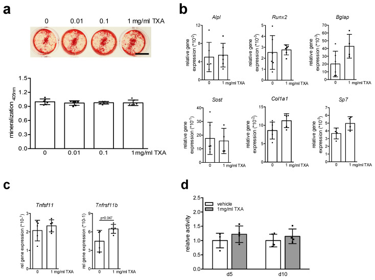
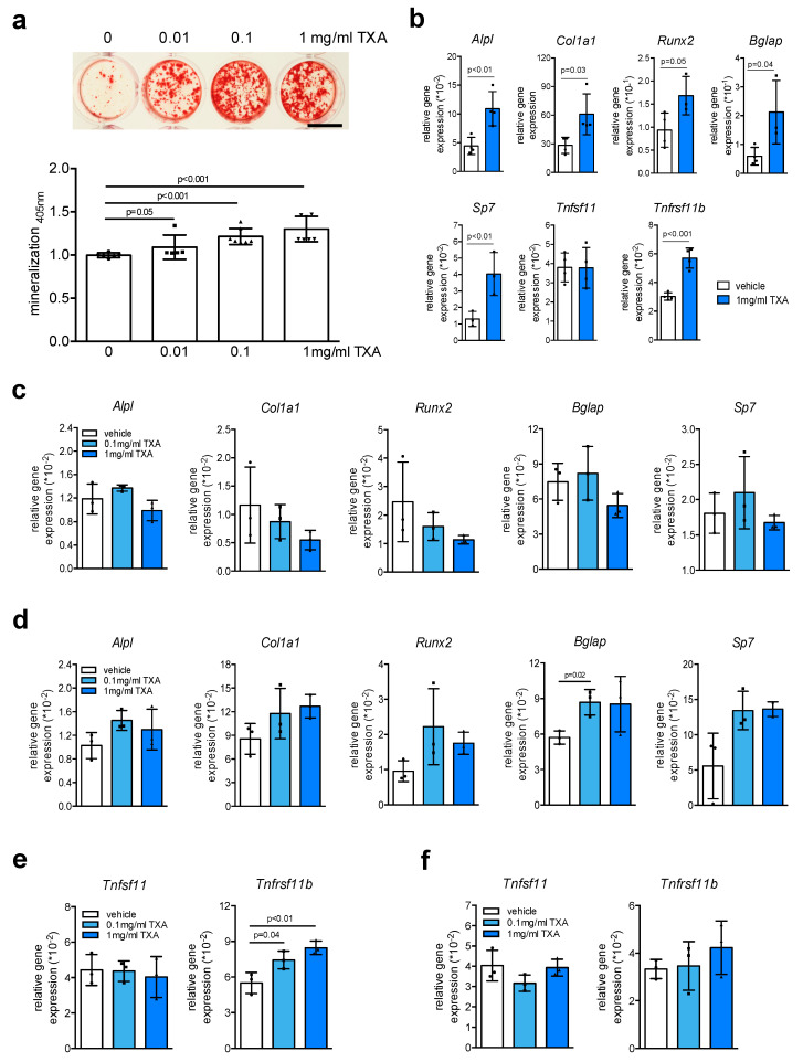
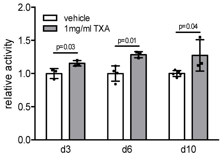
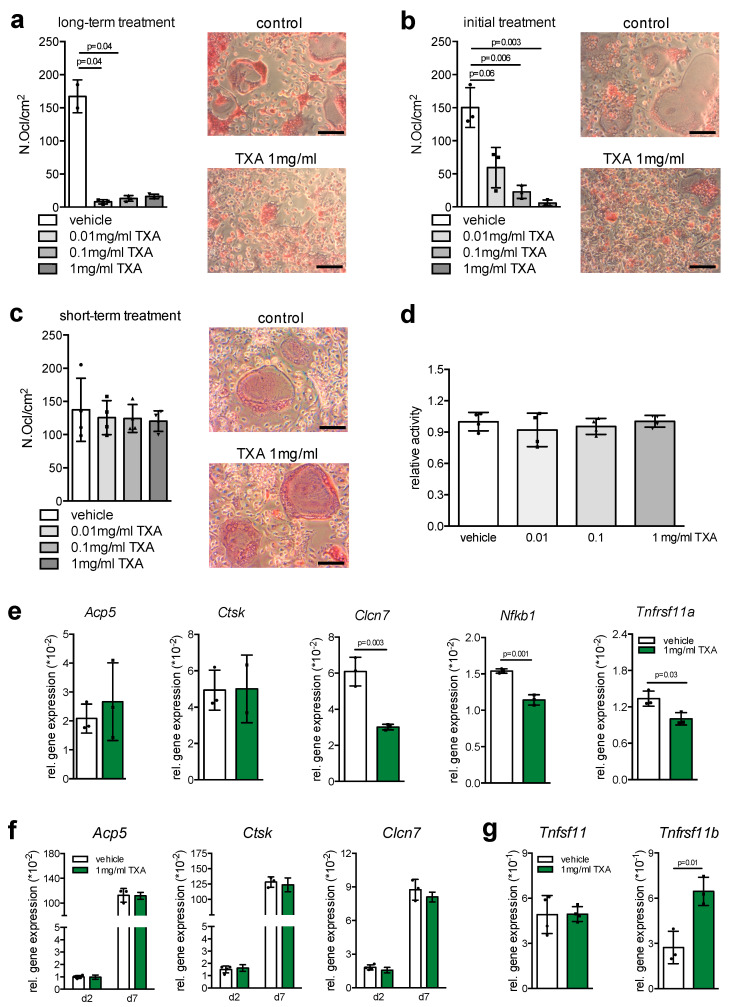
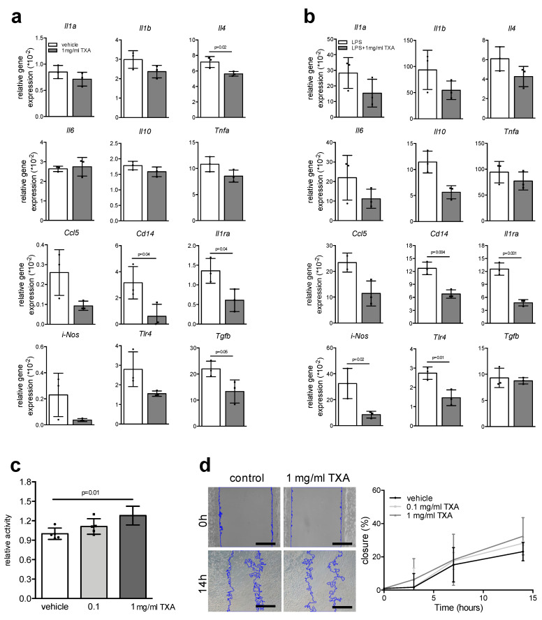
Despite modern surgical trauma care, bleeding contributes to one-third of trauma-related death. A significant improvement was obtained through the introduction of tranexamic acid (TXA), which today is widely used in emergency and elective orthopedic surgery to control bleeding. However, concerns remain regarding potential adverse effects on bone turnover and regeneration. Therefore, we employed standardized cell culture systems including primary osteoblasts, osteoclasts, and macrophages to evaluate potential effects of TXA on murine bone cells. While osteoblasts derived from calvarial digestion were not affected, TXA increased cell proliferation and matrix mineralization in bone marrow-derived osteoblasts. Short-term TXA treatment (6 h) failed to alter the expression of osteoblast markers; however, long-term TXA stimulation (10 days) was associated with the increased expression of genes involved in osteoblast differentiation and extracellular matrix synthesis. Similarly, whereas short-term TXA treatment did not affect gene expression in terminally differentiated osteoclasts, long-term TXA stimulation resulted in the potent inhibition of osteoclastogenesis. Finally, in bone marrow-derived macrophages activated with LPS, simultaneous TXA treatment led to a reduced expression of inflammatory cytokines and chemokines. Collectively, our study demonstrates a differential action of TXA on bone cells including osteoanabolic, anti-resorptive, and anti-inflammatory effects in vitro which suggests novel treatment applications.
Keywords: bone regeneration; macrophages; osteoblast; osteoclast; tranexamic acid.
Conflict of interest statement
The authors declare no conflict of interest.
Figures
Similar articles
-
Interleukin-33, a target of parathyroid hormone and oncostatin m, increases osteoblastic matrix mineral deposition and inhibits osteoclast formation in vitro.Endocrinology. 2011 May;152(5):1911-22. doi: 10.1210/en.2010-1268. Epub 2011 Mar 1. Endocrinology. 2011. PMID: 21363931
-
Inhibition of osteoclast differentiation and collagen antibody-induced arthritis by CTHRC1.Bone. 2017 Apr;97:153-167. doi: 10.1016/j.bone.2017.01.022. Epub 2017 Jan 21. Bone. 2017. PMID: 28115279 Free PMC article.
-
Imatinib promotes osteoblast differentiation by inhibiting PDGFR signaling and inhibits osteoclastogenesis by both direct and stromal cell-dependent mechanisms.J Bone Miner Res. 2007 Nov;22(11):1679-89. doi: 10.1359/jbmr.070719. J Bone Miner Res. 2007. PMID: 17663639
-
Cellular and molecular effects of growth hormone and estrogen on human bone cells.APMIS Suppl. 1997;71:1-30. APMIS Suppl. 1997. PMID: 9357492 Review.
-
Lysophosphatidic acid: a potential mediator of osteoblast-osteoclast signaling in bone.Biochim Biophys Acta. 2013 Jan;1831(1):109-16. doi: 10.1016/j.bbalip.2012.08.001. Epub 2012 Aug 7. Biochim Biophys Acta. 2013. PMID: 22892679 Review.
Cited by
-
Cytotoxicity of tranexamic acid to tendon and bone in vitro: Is there a safe dosage?J Orthop Surg Res. 2022 May 15;17(1):273. doi: 10.1186/s13018-022-03167-5. J Orthop Surg Res. 2022. PMID: 35570313 Free PMC article.
-
The emerging role of tranexamic acid and its principal target, plasminogen, in skeletal health.Acta Pharm Sin B. 2024 Jul;14(7):2869-2884. doi: 10.1016/j.apsb.2024.03.033. Epub 2024 Mar 30. Acta Pharm Sin B. 2024. PMID: 39027253 Free PMC article. Review.
-
Tranexamic Acid Attenuates the Progression of Posttraumatic Osteoarthritis in Mice.Am J Sports Med. 2024 Mar;52(3):766-778. doi: 10.1177/03635465231220855. Epub 2024 Feb 2. Am J Sports Med. 2024. PMID: 38305280 Free PMC article.
-
Evaluation of Novel Tranexamic Acid/Montmorillonite Intercalation Composite, as a New Type of Hemostatic Material.Biomed Res Int. 2022 Feb 28;2022:3963681. doi: 10.1155/2022/3963681. eCollection 2022. Biomed Res Int. 2022. Retraction in: Biomed Res Int. 2023 Nov 29;2023:9782085. doi: 10.1155/2023/9782085. PMID: 35265711 Free PMC article. Retracted.
-
Bidirectional Interaction Between the Brain and Bone in Traumatic Brain Injury.Adv Sci (Weinh). 2025 Aug;12(31):e03149. doi: 10.1002/advs.202503149. Epub 2025 Jul 14. Adv Sci (Weinh). 2025. PMID: 40657694 Free PMC article. Review.
References
-
- Rossaint R., Bouillon B., Cerny V., Coats T.J., Duranteau J., Fernandez-Mondejar E., Filipescu D., Hunt B.J., Komadina R., Nardi G., et al. The European guideline on management of major bleeding and coagulopathy following trauma: Fourth edition. Crit. Care. 2016;20:100. doi: 10.1186/s13054-016-1265-x. - DOI - PMC - PubMed
MeSH terms
Substances
Grants and funding
LinkOut - more resources
Full Text Sources
Other Literature Sources
