. 2021 Jan 5;11(1):54.
doi: 10.3390/brainsci11010054.

Individual Differences in Hemodynamic Responses Measured on the Head Due to a Long-Term Stimulation Involving Colored Light Exposure and a Cognitive Task: A SPA-fNIRS Study

Affiliations

Individual Differences in Hemodynamic Responses Measured on the Head Due to a Long-Term Stimulation Involving Colored Light Exposure and a Cognitive Task: A SPA-fNIRS Study

Hamoon Zohdi et al. Brain Sci. .

Abstract

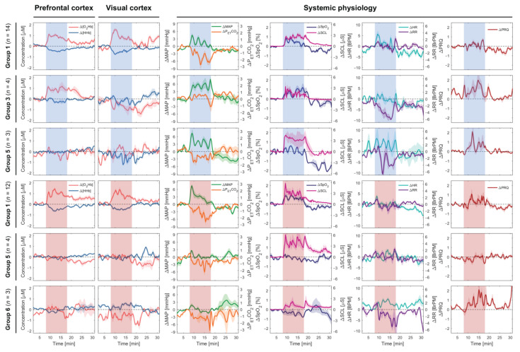
When brain activity is measured by neuroimaging, the canonical hemodynamic response (increase in oxygenated hemoglobin ([O2Hb]) and decrease in deoxygenated hemoglobin ([HHb]) is not always seen in every subject. The reason for this intersubject-variability of the responses is still not completely understood. This study is performed with 32 healthy subjects, using the systemic physiology augmented functional near-infrared spectroscopy (SPA-fNIRS) approach. We investigate the intersubject variability of hemodynamic and systemic physiological responses, due to a verbal fluency task (VFT) under colored light exposure (CLE; blue and red). Five and seven different hemodynamic response patterns were detected in the subgroup analysis of the blue and red light exposure, respectively. We also found that arterial oxygen saturation and mean arterial pressure were positively correlated with [O2Hb] at the prefrontal cortex during the CLE-VFT independent of the color of light and classification of the subjects. Our study finds that there is substantial intersubject-variability of cerebral hemodynamic responses, which is partially explained by subject-specific systemic physiological changes induced by the CLE-VFT. This means that both subgroup analyses and the additional assessment of systemic physiology are of crucial importance to achieve a comprehensive understanding of the effects of a CLE-VFT on human subjects.

Keywords: SPA-fNIRS; cerebral hemodynamics; colored light exposure; laterality; systemic physiology; systemic physiology augmented functional near-infrared spectroscopy; verbal fluency task.


Conflict of interest statement

The authors declare no potential conflicts of interest with respect to the research, authorship, and/or publication of this article.

Figures

Figure 1
Figure 1
(a) Schematic illustration of the measurement protocol; (b) visualization of the placement of devices and sensors on the subject; (c) experimental setup with the position of the subject, illuminated screen, and spotlights. Two colors, i.e., either red or blue were used for the light exposure; in (a,c), the case of blue light exposure is visualized.
Figure 2
Figure 2
Subgroup analysis (the first three common patterns) of cerebral hemodynamics and systemic physiological parameters evoked by the CLE (blue vs. red light) and VFT. The red and blue shaded areas represent the task/stimulation periods during which the subjects were exposed to the respective colors. Median ± standard error of median (SEM) are shown.
Figure 3
Figure 3
Scatter plots displaying Δ[O2Hb] at the PFC and VC vs. other systemic physiological parameters during the CLE-VFT phase at the individual level independent of the color of light. The linear fit is presented for pairs with a significant correlation. The grey shaded areas show 95% of confidence intervals.
Figure 4
Figure 4
Evoked changes of StO2 at the left and right (a) VC and (b) PFC for the blue and red light exposure during the CLE-VFT; (c) Effects of the CLE on the task performance of the subjects. The asterisks indicate the level of significance (p < 0.05, Wilcoxon signed-rank test).

References

    1. Vandewalle G., Maquet P., Dijk D.J. Light as a modulator of cognitive brain function. Trends Cogn. Sci. 2009;13:429–438. doi: 10.1016/j.tics.2009.07.004. - DOI - PubMed
    1. Prayag A., Münch M., Aeschbach D., Chellappa S., Gronfier C. Light Modulation of Human Clocks, Wake, and Sleep. Clocks Sleep. 2019;1:193–208. doi: 10.3390/clockssleep1010017. - DOI - PMC - PubMed
    1. Provencio I., Jiang G., Willem J., Hayes W.P., Rollag M.D. Melanopsin: An opsin in melanophores, brain, and eye. Proc. Natl. Acad. Sci. USA. 1998;95:340–345. doi: 10.1073/pnas.95.1.340. - DOI - PMC - PubMed
    1. Provencio I., Rodriguez I.R., Jiang G., Hayes W.P., Moreira E.F., Rollag M.D. A novel human opsin in the inner retina. J. Neurosci. 2000;20:600–605. doi: 10.1523/JNEUROSCI.20-02-00600.2000. - DOI - PMC - PubMed
    1. Bailes H.J., Lucas R.J. Human melanopsin forms a pigment maximally sensitive to blue light (λmax ≈ 479 nm) supporting activation of Gq/11 and Gi/o signalling cascades. Proc. R. Soc. B Biol. Sci. 2013;280:20122987. doi: 10.1098/rspb.2012.2987. - DOI - PMC - PubMed

LinkOut - more resources