Review
. 2021 Jan 5;22(1):469.
doi: 10.3390/ijms22010469.

Intracellular Ca2+ Signaling in Protozoan Parasites: An Overview with a Focus on Mitochondria

Affiliations
Review

Intracellular Ca2+ Signaling in Protozoan Parasites: An Overview with a Focus on Mitochondria

Pedro H Scarpelli et al. Int J Mol Sci. .

Abstract

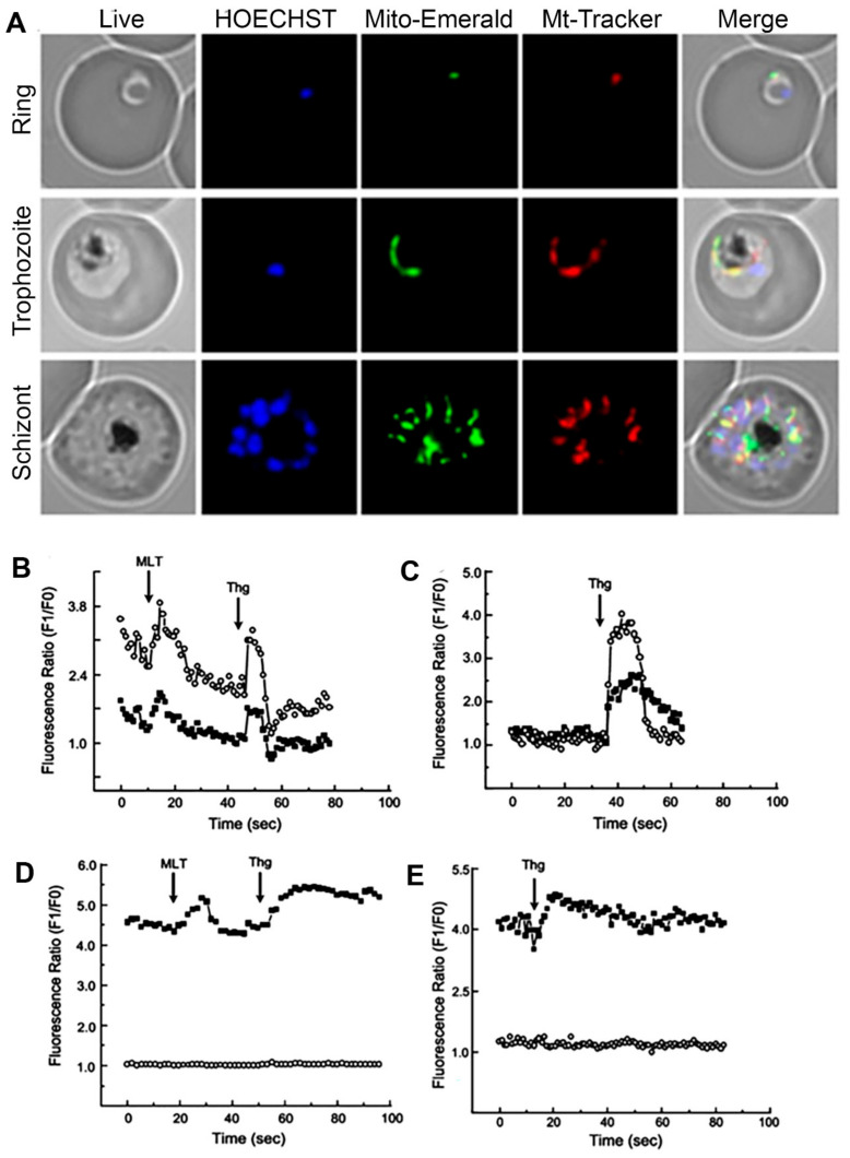
Ca2+ signaling has been involved in controling critical cellular functions such as activation of proteases, cell death, and cell cycle control. The endoplasmatic reticulum plays a significant role in Ca2+ storage inside the cell, but mitochondria have long been recognized as a fundamental Ca2+ pool. Protozoan parasites such as Plasmodium falciparum, Toxoplasma gondii, and Trypanosoma cruzi display a Ca2+ signaling toolkit with similarities to higher eukaryotes, including the participation of mitochondria in Ca2+-dependent signaling events. This review summarizes the most recent knowledge in mitochondrial Ca2+ signaling in protozoan parasites, focusing on the mechanism involved in mitochondrial Ca2+ uptake by pathogenic protists.

Keywords: calcium signaling; mitochondria; protozoan parasites.


Conflict of interest statement

The authors declare no conflict of interest.

Figures

Figure 1
Figure 1
Fluorescent microscopy Plasmodium falciparum ring, trophozoites, and schizonts stages using a mitochondrial green fluorescent protein (GFP) construction: the parasite nucleus was stained by HOECHST33342 (blue) and mitochondria with MitoTracker Red CMX Ros (red) to demonstrate co-localization with Mito-Emerald-GFP (green) (A). P. chabaudi parasites were stained with Fluo4 (cytoplasmatic calcium indicator) and Rhod2 (mitochondrial calcium indicator): effect of melatonin (MLT) and thapsigargin (Thg) on Ca2+ (B); addition of thapsigargin (C); effect of melatonin and thapsigargin on Ca2+ fluorescence in the presence of FCCP (D); addition of thapsigargin in the presence of FCCP (E). Traces represent fluorescence intensity ratio of Ca2+ probes Rhod-2 AM-mitochondria (open circles) and Fluo-3 AM-cytosol (fill squares). The images were retrieved with the author’s consent from the following references: [84] (A) and [83] (BE).

References

    1. Berridge M.J., Bootman M.D., Roderick H.L. Calcium signalling: Dynamics, homeostasis and remodelling. Nat. Rev. Mol. Cell Biol. 2003;4:517–529. doi: 10.1038/nrm1155. - DOI - PubMed
    1. Dupont G., Combettes L., Bird G.S., Putney J.W. Calcium oscillations. Cold Spring Harb. Perspect. Biol. 2011;3:a004226. doi: 10.1101/cshperspect.a004226. - DOI - PMC - PubMed
    1. Plattner H., Verkhratsky A. Ca2+ signalling early in evolution--all but primitive. J. Cell Sci. 2013;126:2141–2150. doi: 10.1242/jcs.127449. - DOI - PubMed
    1. Rizzuto R., De Stefani D., Raffaello A., Mammucari C. Mitochondria as sensors and regulators of calcium signalling. Nat. Rev. Mol. Cell Biol. 2012;13:566–578. doi: 10.1038/nrm3412. - DOI - PubMed
    1. Parekh A.B., Putney J.W. Store-operated calcium channels. Physiol. Rev. 2005;85:757–810. doi: 10.1152/physrev.00057.2003. - DOI - PubMed

LinkOut - more resources