. 2021 Jan 5;22(1):465.
doi: 10.3390/ijms22010465.

Prion-Associated Neurodegeneration Causes Both Endoplasmic Reticulum Stress and Proteasome Impairment in a Murine Model of Spontaneous Disease

Affiliations

Prion-Associated Neurodegeneration Causes Both Endoplasmic Reticulum Stress and Proteasome Impairment in a Murine Model of Spontaneous Disease

Alicia Otero et al. Int J Mol Sci. .

Abstract

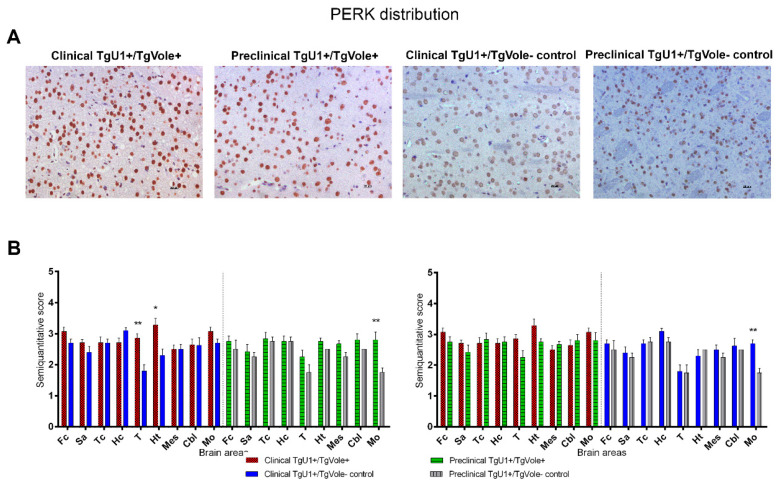
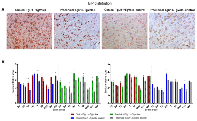
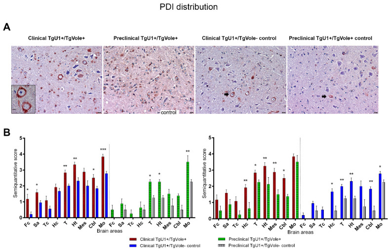
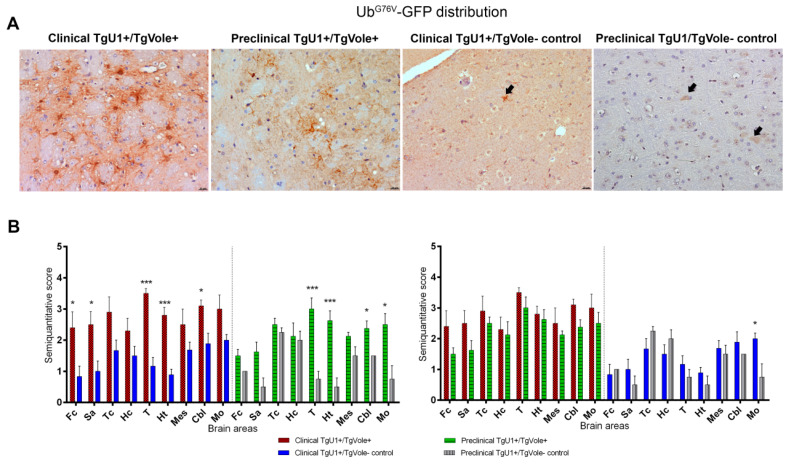
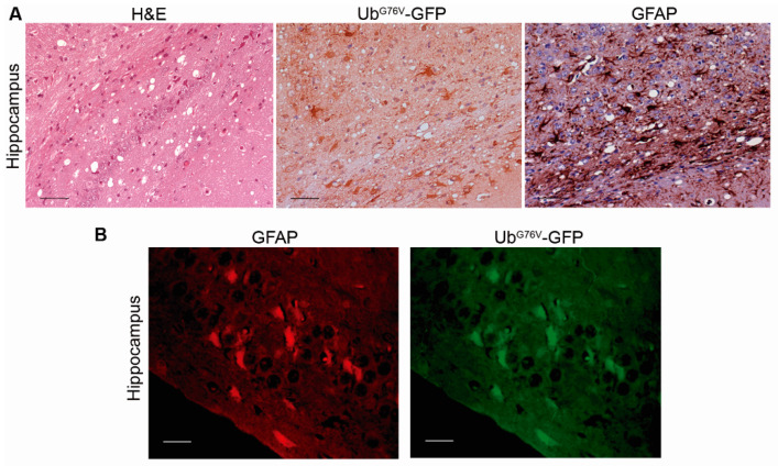
Prion diseases are a group of neurodegenerative disorders that can be spontaneous, familial or acquired by infection. The conversion of the prion protein PrPC to its abnormal and misfolded isoform PrPSc is the main event in the pathogenesis of prion diseases of all origins. In spontaneous prion diseases, the mechanisms that trigger the formation of PrPSc in the central nervous system remain unknown. Several reports have demonstrated that the accumulation of PrPSc can induce endoplasmic reticulum (ER) stress and proteasome impairment from the early stages of the prion disease. Both mechanisms lead to an increment of PrP aggregates in the secretory pathway, which could explain the pathogenesis of spontaneous prion diseases. Here, we investigate the role of ER stress and proteasome impairment during prion disorders in a murine model of spontaneous prion disease (TgVole) co-expressing the UbG76V-GFP reporter, which allows measuring the proteasome activity in vivo. Spontaneously prion-affected mice showed a significantly higher accumulation of the PKR-like ER kinase (PERK), the ER chaperone binding immunoglobulin protein (BiP/Grp78), the ER protein disulfide isomerase (PDI) and the UbG76V-GFP reporter than age-matched controls in certain brain areas. The upregulation of PERK, BiP, PDI and ubiquitin was detected from the preclinical stage of the disease, indicating that ER stress and proteasome impairment begin at early stages of the spontaneous disease. Strong correlations were found between the deposition of these markers and neuropathological markers of prion disease in both preclinical and clinical mice. Our results suggest that both ER stress and proteasome impairment occur during the pathogenesis of spontaneous prion diseases.

Keywords: ER stress; UPS impairment; endoplasmic reticulum; prions; proteasome.


Conflict of interest statement

The authors declare no conflict of interest.

Figures

Figure 1
Figure 1
PRKR-like endoplasmic reticulum kinase (PERK) upregulation in the thalamic area of clinical TgU1+/TgVole+ mice. (A) Nuclear staining was observed for PERK in numerous cells of all groups of mice. The number of immunopositive cells was higher and immunostaining was stronger in TgU1+/TgVole+ mice, especially in the thalamus and hypothalamus. Images correspond to the hypothalamic area in all mice. (B) PERK distribution in the brains of clinical and preclinical TgU1+/TgVole+ mice and their age-matched controls. PERK immunostaining was analyzed using a semiquantitative scale from 0 (lack of immunostaining) to 5 (very intense immunostaining) in nine different brain areas: frontal cortex (Fc), septal area (Sa), cortex at the level of the thalamus (Tc), hippocampus (Hc), thalamus (T), hypothalamus (Ht), mesencephalon (Mes), cerebellum (Cbl) and medulla oblongata (Mo). The number of animals studied was the following: clinical TgU1+/TgVole+ n = 6 (4 female, 2 male), clinical TgU1+/TgVole controls n = 8 (6 female, 2 male), preclinical TgU1+/TgVole+ n = 5 (2 female, 3 male) and preclinical TgU1+/TgVole controls n = 5 (2 female, 3 male). Comparison of the PERK immunolabeling revealed significant differences between clinical TgU1+/TgVole+ mice and clinical TgU1+/TgVole controls and between preclinical TgU1+/TgVole+ mice and preclinical TgU1+/TgVole controls in different brain areas. (* p < 0.05, ** p < 0.01, Mann-Whitney U test).
Figure 2
Figure 2
Binding immunoglobulin protein (BiP) expression levels are higher in clinical and preclinical TgU1+/TgVole+ mice than in healthy TgU1+/TgVole- controls in certain brain areas. (A) All groups of mice presented uniform cytoplasmic labeling of BiP in numerous neurons and a diffuse staining in the neuropil. Immunostaining intensity, rather than the number of positive cells, was higher in both clinical and preclinical TgU1+/TgVole+ compared to their age-matched controls. (B) BiP distribution in the brains of clinical and preclinical TgU1+/TgVole+ mice and their age-matched controls. BiP immunostaining was analyzed using a semiquantitative scale from 0 (lack of immunostaining) to 5 (very intense immunostaining) in nine different brain areas: frontal cortex (Fc), septal area (Sa), cortex at the level of the thalamus (Tc), hippocampus (Hc), thalamus (T), hypothalamus (Ht), mesencephalon (Mes), cerebellum (Cbl) and medulla oblongata (Mo). The number of animals included in each group was: clinical TgU1+/TgVole+ n = 6 (4 female, 2 male), clinical TgU1+/TgVole controls n = 8 (6 female, 2 male), preclinical TgU1+/TgVole+ n = 5 (2 female, 3 male) and preclinical TgU1+/TgVole controls n = 5 (2 female, 3 male). Comparison of the BiP immunolabeling revealed significant differences between clinical TgU1+/TgVole+ mice and clinical TgU1+/TgVole controls and between preclinical TgU1+/TgVole+ mice and preclinical TgU1+/TgVole controls in different brain areas. No differences were observed between clinical and preclinical TgU1+/TgVole+ mice. (* p < 0.05, ** p < 0.01, Mann-Whitney U test).
Figure 3
Figure 3
Protein disulfide isomerase (PDI) accumulation is more intense in clinical TgU1+/TgVole+ mice than in preclinical TgU1+/TgVole+ mice and healthy TgU1+/TgVole controls in all brain areas. (A) A strong intraneuronal PDI labeling was observed in the gigantocellular reticular nucleus of the medulla oblongata of clinical TgU1+/TgVole+ mice. Intraneuronal PDI immunolabeling was also observed in the other groups of mice, but the intensity of the immunostaining and the number of immunopositive cells were reduced compared with clinical TgU1+/TgVole+ mice (arrows). Insert picture contains two neurons showing a strong accumulation of PDI. (B) PDI distribution in the brains of clinical and preclinical TgU1+/TgVole+ mice and their age-matched controls. PDI immunolabeling was semiquantitatively analyzed and scored on a scale of 0 (absence of immunolabeling) to 5 (very intense immunolabeling) in nine brain areas. Each group included the following number of animals: clinical TgU1+/TgVole+ n = 6 (4 female, 2 male), clinical TgU1+/TgVole controls n = 8 (6 female, 2 male), preclinical TgU1+/TgVole+ n = 5 (2 female, 3 male) and preclinical TgU1+/TgVole controls n = 5 (2 female, 3 male). Comparison of the PDI immunolabeling profiles revealed significant differences between the group of clinical TgU1+/TgVole+ mice and clinical TgU1+/TgVole controls in certain brain areas and between preclinical TgU1+/TgVole+ mice and preclinical TgU1+/TgVole controls. Significant differences in PDI immunostaining were also noticed between clinical and preclinical TgU1+/TgVole+ mice and between clinical and preclinical TgU1+/TgVole controls. (* p < 0.05, ** p < 0.01, *** p < 0.001, Mann-Whitney U test).
Figure 4
Figure 4
UbG76V-GFP accumulation is more intense in clinical and preclinical TgU1+/TgVole+ mice than in healthy TgU1+/TgVole controls in certain brain areas. (A) Strong UbG76V-GFP immunostaining was observed in the thalamus of clinical and preclinical TgU1+/TgVole+ mice, affecting cells whose morphology is compatible with reactive astrocytes. Healthy aged TgU1+/TgVole mice (clinical TgU1+/TgVole controls) showed granular immunostaining and filamentous ubiquitin aggregates in the neuropil (arrow). Healthy young TgU1+/TgVole mice (preclinical TgU1+/TgVole controls) showed slight intraneuronal UbG76V-GFP immunolabeling (arrows). (B) UbG76V-GFP distribution in the brains of clinical and preclinical TgU1+/TgVole+ mice and their age-matched controls. UbG76V-GFP immunolabeling was semiquantitatively analyzed and scored on a scale of 0 (absence of immunolabeling) to 5 (very intense immunolabeling) in nine different brain areas. The number of animals within each group was: clinical TgU1+/TgVole+ n = 6 (4 female, 2 male), clinical TgU1+/TgVole controls n = 8 (6 female, 2 male), preclinical TgU1+/TgVole+ n = 5 (2 female, 3 male) and preclinical TgU1+/TgVole controls n = 5 (2 female, 3 male). Comparison of the UbG76V-GFP immunolabeling profiles revealed significant differences between the group of clinical TgU1+/TgVole+ mice and clinical TgU1+/TgVole controls and between preclinical TgU1+/TgVole+ mice and preclinical TgU1+/TgVole controls in numerous brain areas. No differences were observed between clinical and preclinical TgU1+/TgVole+ mice. (* p < 0.05, *** p < 0.001, Mann-Whitney U test).
Figure 5
Figure 5
UbG76V-GFP intracellular accumulation is observed in brain areas showing prion-associated neuropathology. (A) Hippocampus from a clinical TgU1+/TgVole+ mouse stained with hematoxylin and eosin and immunostained for UbG76V-GFP and glial fibrillary acidic protein (GFAP). This animal shows severe spongiosis in the hippocampus and intense immunostaining for UbG76V-GFP and GFAP. Strong UbG76V-GFP immunolabeling is observed, affecting numerous cells that appear to be reactive astrocytes. (B) Dual immunofluorescence staining with anti-GFAP and anti-GFP antibodies revealed that numerous reactive astrocytes accumulated the UbG76V-GFP reporter, suggesting that these cells can compensate for proteasome impairment and accumulate high amounts of ubiquitin conjugates before succumbing to the cytotoxic effect.

References

    1. Prusiner S.B. Prions. Proc. Natl. Acad. Sci. USA. 1998;95:13363–13383. doi: 10.1073/pnas.95.23.13363. - DOI - PMC - PubMed
    1. Collinge J. Prion Diseases of Humans and Animals: Their Causes and Molecular Basis. Annu. Rev. Neurosci. 2001;24:519–550. doi: 10.1146/annurev.neuro.24.1.519. - DOI - PubMed
    1. Hetz C., Glimcher L.H. Fine-Tuning of the Unfolded Protein Response: Assembling the IRE1α Interactome. Mol. Cell. 2009;35:551–561. doi: 10.1016/j.molcel.2009.08.021. - DOI - PMC - PubMed
    1. Soto C., Satani N. The intricate mechanisms of neurodegeneration in prion diseases. Trends Mol. Med. 2011;17:14–24. doi: 10.1016/j.molmed.2010.09.001. - DOI - PMC - PubMed
    1. Torres M., Castillo K., Armisén R., Stutzin A., Soto C., Hetz C. Prion Protein Misfolding Affects Calcium Homeostasis and Sensitizes Cells to Endoplasmic Reticulum Stress. PLoS ONE. 2010;5:e15658. doi: 10.1371/journal.pone.0015658. - DOI - PMC - PubMed

Grants and funding