. 2021 Jan 5;9(1):41.
doi: 10.3390/biomedicines9010041.

Photobiomodulation of Human Fibroblasts and Keratinocytes with Blue Light: Implications in Wound Healing

Affiliations

Photobiomodulation of Human Fibroblasts and Keratinocytes with Blue Light: Implications in Wound Healing

Francesca Rossi et al. Biomedicines. .

Abstract

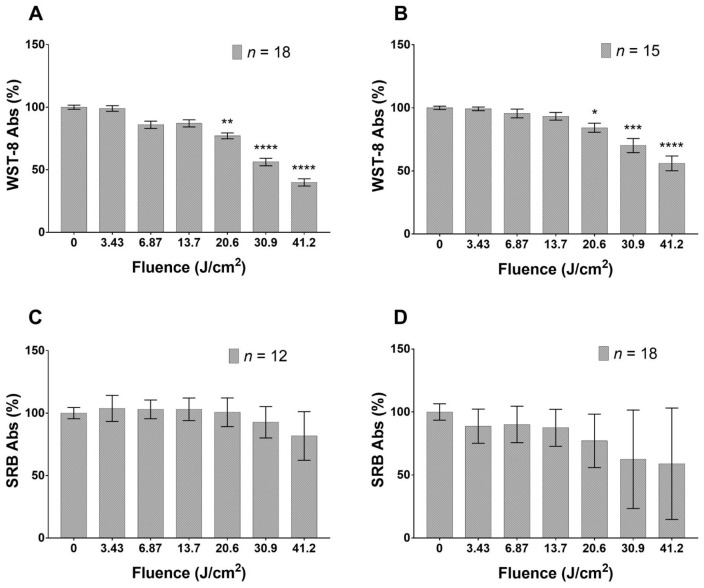
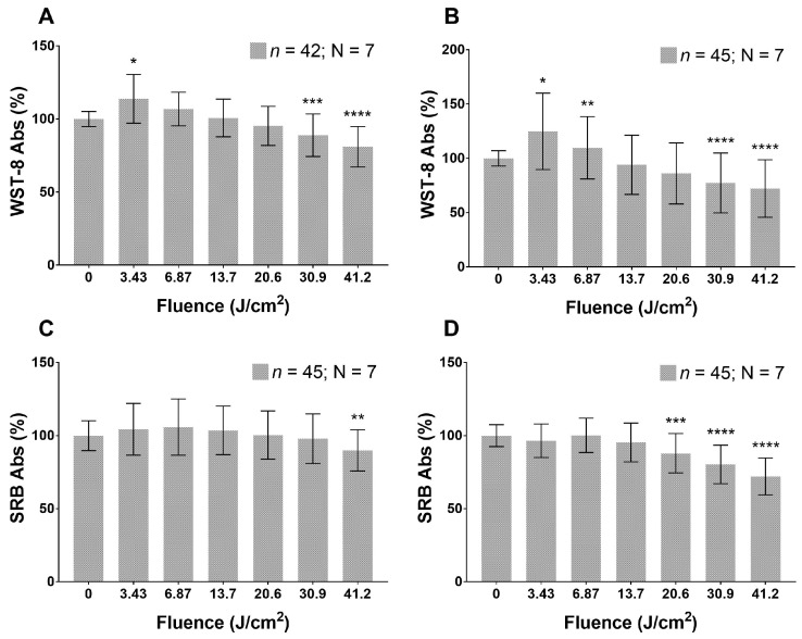
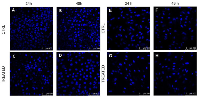
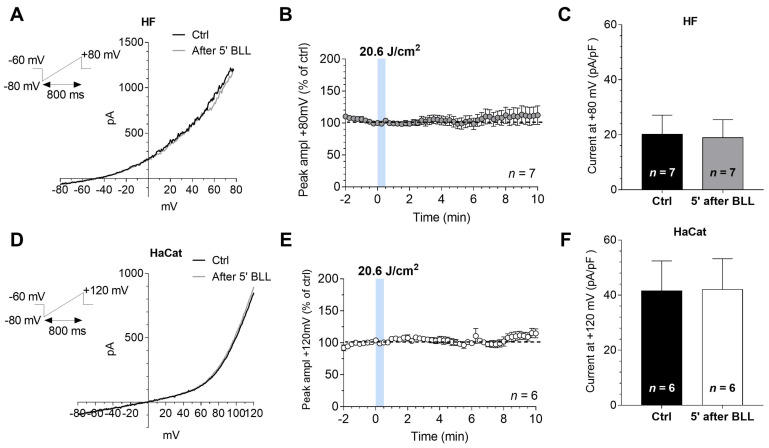
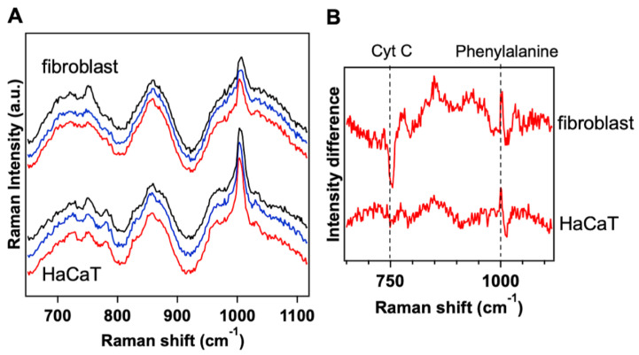
In recent years, photobiomodulation (PBM) has been recognized as a physical therapy in wound management. Despite several published research papers, the mechanism underlying photobiomodulation is still not completely understood. The investigation about application of blue light to improve wound healing is a relatively new research area. Tests in selected patients evidenced a stimulation of the healing process in superficial and chronic wounds treated with a blue LED light emitting at 420 nm; a study in animal model pointed out a faster healing process in superficial wound, with an important role of fibroblasts and myofibroblasts. Here, we present a study aiming at evidencing the effects of blue light on the proliferation and metabolism in fibroblasts from healthy skin and keratinocytes. Different light doses (3.43, 6.87, 13.7, 20.6, 30.9 and 41.2 J/cm2) were used to treat the cells, evidencing inhibitory and stimulatory effects following a biphasic dose behavior. Electrophysiology was used to investigate the effects on membrane currents: healthy fibroblasts and keratinocytes showed no significant differences between treated and not treated cells. Raman spectroscopy revealed the mitochondrial Cytochrome C (Cyt C) oxidase dependence on blue light irradiation: a significant decrease in peak intensity of healthy fibroblast was evidenced, while it is less pronounced in keratinocytes. In conclusion, we observed that the blue LED light can be used to modulate metabolism and proliferation of human fibroblasts, and the effects in wound healing are particularly evident when studying the fibroblasts and keratinocytes co-cultures.

Keywords: LED; blue light; photobiomodulation; wound healing.


Conflict of interest statement

The authors declare no conflict of interest. The funders had no role in the design of the study and in the decision to publish the results.

Figures

Figure 1
Figure 1
Effects of blue LED light on metabolism and proliferation of HaCaT cells: (A,B) cell metabolism 24 and 48 h after treatment, respectively; and (C,D) proliferation 24 and 48 h after treatment, respectively. Data are expressed as mean ± SD. Each measure is repeated in duplicate for each condition. Statistical analysis: * p < 0.05; ** p < 0.01; *** p < 0.001; **** p < 0.0001 vs. control (not irradiated cells), Kruskal–Wallis test followed by Dunn’s post-hoc test.
Figure 2
Figure 2
Effects of blue LED light on metabolism and proliferation of fibroblasts cells: (A,B) metabolism 24 and 48 h after treatment, respectively; and (C,D) proliferation 24 and 48 h after treatment. Data are expressed as mean ± SD. Each measure is repeated in triplicate; n, number of replicates; N, number of human samples. Statistical analysis: * p < 0.05; ** p < 0.01; *** p < 0.001; **** p < 0.0001 vs. control (not irradiated cells), Kruskal–Wallis test followed by Dunn’s post-hoc test.
Figure 3
Figure 3
DAPI staining performed on cultured human fibroblasts (right) and HaCaT cells (left) shows differences in treated and control samples: (A,B) control HaCaT samples analyzed 24 and 48 h after the beginning of the experiment; (C,D) treated HaCaT cells analyzed at 24 and 48 h after the application of blue light with 41.2 J/cm2 fluence; (E,F) control fibroblasts samples analyzed 24 and 48 h after the beginning of the experiment; and (G,H) treated fibroblasts cells analyzed at 24 and 48 h after treatment with a blue light dose of 41.2 J/cm2, respectively. Cells nuclei are in blue.
Figure 4
Figure 4
The application of blue LED light does not modify ramp-evoked currents in both cultured fibroblasts and HaCat cells: (A) original whole-cell patch clamp current traces evoked by a voltage ramp protocol (from 80 to +80 mV, 800 ms, top left inset) before (Ctrl, black trace) or 5 min after the application of 20.6 J/cm2 blue LED light (BLL, grey trace) in a typical fibroblast (HF); (B) averaged time courses (mean ± SEM) of ramp-evoked currents at +80 mV, expressed as percent of ctrl, in HFs (n = 7) before, during or after the application of blue LED light; (C) pooled data (mean ± SEM) of ramp current amplitude at +80 mV, recorded before or after 5 min of blue LED light (BLL) application in HFs (n = 7); (D) original whole-cell patch clamp current traces evoked by a voltage ramp protocol (from 80 to +120 mV, 800 ms, top left inset) before (Ctrl, black trace) or 5 min after the application of 20.6 J/cm2 blue LED light (BLL, grey trace) in a typical HaCat cell; (E) averaged time courses (mean ± SEM) of ramp-evoked currents at +120 mV, expressed as percent of ctrl, in HaCat cells (n = 6) before, during or after the application of 20.6 J/cm2 fluence of blue LED light; and (F) pooled data (mean ± SEM) of ramp current amplitude at +120 mV, recorded before or after 5 min of blue LED light (BLL) application in HaCat (n = 6).
Figure 5
Figure 5
The Raman intensity of Cytochrome C peak at 750 cm1 undergoes significant variation in fibroblast cells and minor variation in HaCaT cells upon blue LED light irradiation: (A) averaged Raman spectra acquired at least on 20 fibroblasts and HaCaT cells before the treatment (black) and after 20.6 J/cm2 (blue) and 41.2 J/cm2 (red) of blue LED light; and (B) differential Raman spectrum obtained by subtracting the spectrum acquired before the treatment with blue light from the spectrum acquired after the application of blue light at 41.2 J/cm2, both for cultured fibroblast and HaCaT cells. Phenylalanine mode at 1003 cm1 was used for normalizing the Raman spectra of the cells.
Figure 6
Figure 6
Blue LED light stimulates in vitro fibroblasts and keratinocytes migration: (A) column bars represent the percentage of scratch area (i.e., mimicking the wound area) in co-cultures observed 24, 48 and 72 h after treatment, in comparison to 0 h.; and (B,C) representative images of an untreated (top) and treated (bottom) co-culture of HaCaT and fibroblast, as observed 72 h after treatment (fluence: 20.6 J/cm2). Data are expressed as mean ± SD, n = 2. Statistical analysis: **** p < 0.0001, t24; t48; t72 vs. t0, one-way ANOVA followed by Dunnett’s multiple comparison test.

References

    1. Piaggesi A., Låuchli S., Bassetto F., Biedermann T., Marques A., Najafi B., Palla I., Scarpa C., Seimetz D., Triulzi I., et al. Advanced therapies in wound management: Cell and tissue based therapies, physical and bio-physical therapies smart and IT based technologies. J. Wound Care. 2018;27:S1–S137. doi: 10.12968/jowc.2018.27.Sup6a.S1. - DOI - PubMed
    1. Mester E., Szende B., Gärtner P. The effect of laser beams on the growth of hair in mice. Radiobiol. Radiother. 1968;9:621. - PubMed
    1. Heiskanen V., Hamblin M.R. Photobiomodulation: Lasers vs. light emitting diodes? Photochem. Photobiol. Sci. 2018;17:1003–1017. doi: 10.1039/C8PP00176F. - DOI - PMC - PubMed
    1. Dompe C., Moncrieff L., Matys J., Grzech-Leśniak K., Kocherova I., Bryja A., Bruska M., Dominiak M., Mozdziak P., Skiba T.H.I., et al. Photobiomodulation—Underlying Mechanism and Clinical Applications. J. Clin. Med. 2020;9:1724. doi: 10.3390/jcm9061724. - DOI - PMC - PubMed
    1. Serrage H., Heiskanen V., Palin W.M., Cooper P.R., Milward M.R., Hadis M., Hamblin M.R. Under the spotlight: Mechanisms of photobiomodulation concentrating on blue and green light. Photochem. Photobiol. Sci. 2019;18:1877–1909. doi: 10.1039/C9PP00089E. - DOI - PMC - PubMed

LinkOut - more resources