Review
. 2021 Jan 14;10(1):150.
doi: 10.3390/cells10010150.

Microglial Turnover in Ageing-Related Neurodegeneration: Therapeutic Avenue to Intervene in Disease Progression

Affiliations
Review

Microglial Turnover in Ageing-Related Neurodegeneration: Therapeutic Avenue to Intervene in Disease Progression

Shofiul Azam et al. Cells. .

Abstract

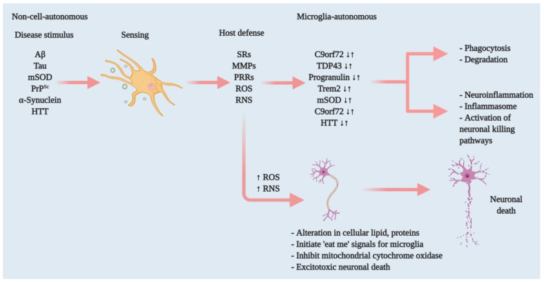
Microglia are brain-dwelling macrophages and major parts of the neuroimmune system that broadly contribute to brain development, homeostasis, ageing and injury repair in the central nervous system (CNS). Apart from other brain macrophages, they have the ability to constantly sense changes in the brain's microenvironment, functioning as housekeepers for neuronal well-being and providing neuroprotection in normal physiology. Microglia use a set of genes for these functions that involve proinflammatory cytokines. In response to specific stimuli, they release these proinflammatory cytokines, which can damage and kill neurons via neuroinflammation. However, alterations in microglial functioning are a common pathophysiology in age-related neurodegenerative diseases, such as Alzheimer's, Parkinson's, Huntington's and prion diseases, as well as amyotrophic lateral sclerosis, frontotemporal dementia and chronic traumatic encephalopathy. When their sentinel or housekeeping functions are severely disrupted, they aggravate neuropathological conditions by overstimulating their defensive function and through neuroinflammation. Several pathways are involved in microglial functioning, including the Trem2, Cx3cr1 and progranulin pathways, which keep the microglial inflammatory response under control and promote clearance of injurious stimuli. Over time, an imbalance in this system leads to protective microglia becoming detrimental, initiating or exacerbating neurodegeneration. Correcting such imbalances might be a potential mode of therapeutic intervention in neurodegenerative diseases.

Keywords: homeostasis; macrophages; microglia; neurodegeneration; neuroinflammation.


Conflict of interest statement

All authors declare no conflict of interest.

Figures

Figure 1
Figure 1
Effectors of microglia function associated with neurodegeneration and how microglia damage or kills neurons. Microglia have both host-defensive and detrimental actions, depending on the scenario. When they encounter aberrant or misfolded proteins—such as Aβ, aggregated α-synuclein, oxidised or mSOD1, or PrPSc—they function as sentinels, protective hosts. In response to these toxic stimuli, microglia attempt to clear aggregated Aβ, α-synuclein or mSOD1 via SRs and other PRRs. However, the nature of the aberrant proteins or their continued production disrupts microglial housekeeping functions. Several pathways activate: NADPH oxidase produces superoxide and derivative oxidants; iNOS produces nitric oxide derivatives, glutamate, cathepsin B and other proteases, and that produces stressed neurons. These processes dysregulate the microglial host-defensive mechanism, leading to an exaggerated proinflammatory response, neurotoxicity and neurodegeneration. Microglia also include tumour necrosis factor (TNF) as an indirect pathway to damage or kill neurons via reducing brain-derived neurotrophic factor (BDNF) and insulin-like growth factor (IGF) production. Neurodegenerative diseases such as AD, ALS and HD, furthermore, cause mutations in specific genes that lead to self-autonomous dysregulation of host defence. This initiates or exaggerates proinflammatory responses, resulting in neurotoxicity and neurodegeneration. In this way, when mutations in TDP-43, progranulin and Trem2 increase (↑), they affect phagocytosis and associated degradation pathways. Similarly, mutations in mSOD and HTT (↑) also affect inflammasome activation and neuronal killing pathways. On the other hand, mutations in C9orf72 can affect both phagocytosis and inflammasome pathways. In normal physiology, a microglia-autonomous mechanism controls the scenario by clearing (↓) mutant or aberrant proteins (adapted from ref. [8]).
Figure 2
Figure 2
Microglial regulation of neuronal networks and CNS homeostasis. By playing a phagocytic role, microglia engulf dead or degenerated neurons via TAM receptor-mediated recognition of GAS6-opsonised cells. Microglia control synaptic plasticity by downregulating AMPA receptor activity through ROS secretion and by modulating BDNF/Trk receptor pathways. Beyond this, microglia can strip synapses through Cx3cr1-Cx3cl1 interactions and microglial major histocompatibility complex II (MHCII) [32]. They can also affect neuronal circuitry via interacting with astrocytes through the TNF-mediated pathway, which releases glutamate and ATP from astrocytes.
Figure 3
Figure 3
Effects of ageing on healthy to proinflammatory microglia. Healthy microglia regulate the release of cytokines, proteases, ROS and RNS and phagocyte foreign bodies. In addition, they clear cell debris and proteins. With advanced ageing, microglia eventually lose function or functions dysregulated by persistent exposure to foreign bodies or mutations. Increasing microglial ROS production releases proteases and cytokines and slows the autophagic process, leading to aberrant protein aggregation and inflammatory response, neuronal death and neurodegenerative disease.

References

    1. Crotti A., Ransohoff R.M. Microglial Physiology and Pathophysiology: Insights from Genome-wide Transcriptional Profiling. Immunity. 2016;44:505–515. doi: 10.1016/j.immuni.2016.02.013. - DOI - PubMed
    1. Ginhoux F., Greter M., Leboeuf M., Nandi S., See P., Gokhan S., Mehler M.F., Conway S.J., Ng L.G., Stanley E.R., et al. Fate mapping analysis reveals that adult microglia derive from primitive macrophages. Science. 2010;330:841–845. doi: 10.1126/science.1194637. - DOI - PMC - PubMed
    1. Sheng J., Ruedl C., Karjalainen K. Most Tissue-Resident Macrophages Except Microglia Are Derived from Fetal Hematopoietic Stem Cells. Immunity. 2015;43:382–393. doi: 10.1016/j.immuni.2015.07.016. - DOI - PubMed
    1. Hashimoto D., Chow A., Noizat C., Teo P., Beasley M.B., Leboeuf M., Becker C.D., See P., Price J., Lucas D., et al. Tissue-resident macrophages self-maintain locally throughout adult life with minimal contribution from circulating monocytes. Immunity. 2013;38:792–804. doi: 10.1016/j.immuni.2013.04.004. - DOI - PMC - PubMed
    1. Pelvig D.P., Pakkenberg H., Stark A.K., Pakkenberg B. Neocortical glial cell numbers in human brains. Neurobiol. Aging. 2008;29:1754–1762. doi: 10.1016/j.neurobiolaging.2007.04.013. - DOI - PubMed

Publication types

LinkOut - more resources