. 2021 Jan 14;10(2):281.
doi: 10.3390/jcm10020281.

Uveitis as an Open Window to Systemic Inflammatory Diseases

Affiliations

Uveitis as an Open Window to Systemic Inflammatory Diseases

Thomas El Jammal et al. J Clin Med. .

Abstract

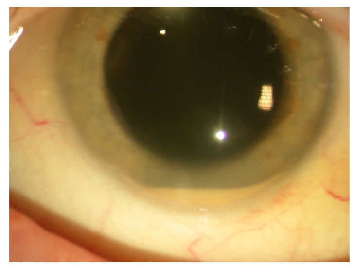
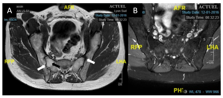
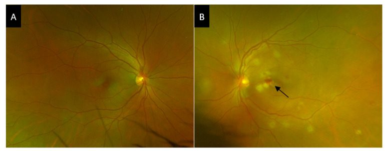
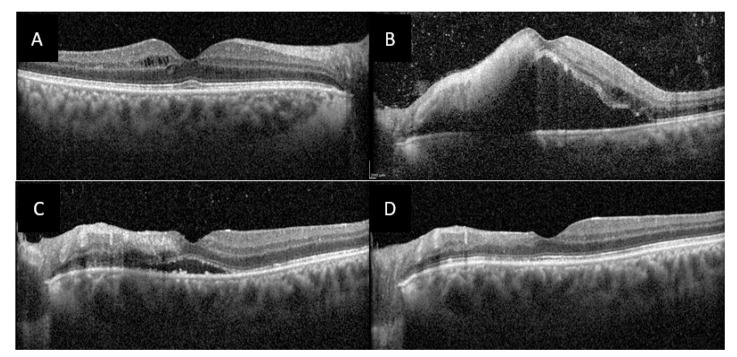
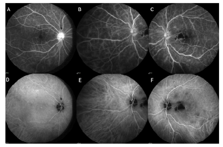
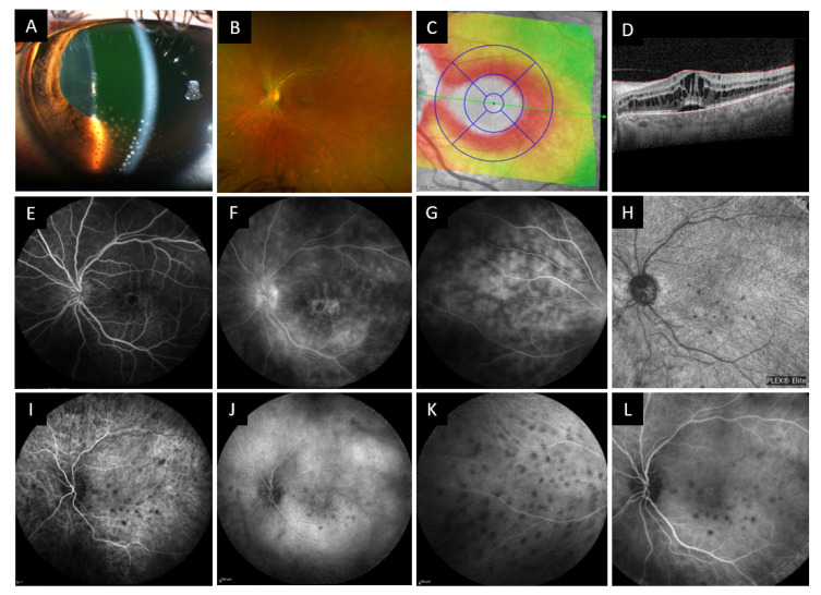
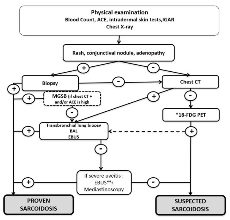
Spondyloarthritis (Spa), Behçet's disease (BD) and sarcoidosis are major systemic inflammatory diseases worldwide. They are all multisystem pathologies and share a possible ocular involvement, especially uveitis. We hereby describe selected cases who were referred by ophthalmologists to our internal medicine department for unexplained uveitis. Physical examination and/or the use of laboratory and imaging investigations allowed to make a diagnosis of a systemic inflammatory disease in a large proportion of patients. In our tertiary referral center, 75 patients have been diagnosed with Spa (n = 20), BD (n = 9), or sarcoidosis (n = 46) in the last two years. There was a significant delay in the diagnosis of Spa-associated uveitis. Screening strategies using Human Leukocyte Antigen (HLA)-B27 determination and sacroiliac magnetic resonance imaging in patients suffering from chronic low back pain and/or psoriasis helped in the diagnosis. BD's uveitis affects young people from both sexes and all origins and usually presents with panuveitis and retinal vasculitis. The high proportion of sarcoidosis in our population is explained by the use of chest computed tomography (CT) and 18F-fluorodeoxyglucose positron emission tomography CT that helped to identify smaller hilar or mediastinal involvement and allowed to further investigate those patients, especially in the elderly. Our results confirm how in these sight- and potentially life-threatening diseases a prompt diagnosis is mandatory and benefits from a multidisciplinary approach.

Keywords: Behçet’s disease; HLA-B27 associated uveitis; sarcoidosis; spondyloarthritis; uveitis.


Conflict of interest statement

Laurent Kodjikian declared conflicts of interest from Abbvie, Allergan, Bayer, Novartis, Roche, Théa. Other authors declare no conflict of interests.

Figures

Figure 1
Figure 1
Slit lamp examination of case 1 showing nongranulomatous corneal precipitates with a cellular anterior chamber reaction and hypopyon.
Figure 2
Figure 2
Axial sacroiliac MRI of case 1. (A): T1-weighted sacroiliac axial MRI of case 1 showing major joint reshaping in the form of hypo dense areas bordering the joint (white arrows) as well as ankylosis of the right sacroiliac joint with a T1 hypersignal of the subchondral bone. (B): T2 SPAIR axial sequence without hypersignal of the sacroiliac joints, indicating the absence of active involvement. Abbreviations: A = anterior, P= posterior, R = right, L = left, H = head, F = foot; MRI: magnetic resonance imaging; T2 SPAIR: T2 Spectral Attenuated Inversion Recovery.
Figure 3
Figure 3
Wide field fundus photograph of the right eye of the case 2 patient. (A), Widefield fundus photograph of the right eye showing no signs of intermediate or posterior uveitis. (B), Widefield fundus photograph of the left eye showing multiple patchy subretinal white spots and a nodule of retinal necrosis with hemorrhage (black arrow).
Figure 4
Figure 4
Optical coherence tomography (OCT) of both macula of case 2 patient. (A), OCT of the right macula showing a few cysts of intraretinal edema. (B), OCT of the left macula showing a large serous retinal detachment, a hyperreflective nodule in the inner retinal layers, intraretinal edema and numerous hyperreflective dots in the vitreous. (C), OCT of the left macula at 1-month follow-up showing partial regression of subretinal fluid. (D) OCT of the left macula at 3 months follow-up showing complete regression of macular edema, and atrophy of internal retina in place of the hyperreflective nodule.
Figure 5
Figure 5
Fluorescein angiogram of both eyes of case 2 patient. Fluorescein angiogram of the right eye (A) in late sequence showing diffuse capillaritis and papillitis. Fluorescein angiogram of the left eye (B,C) in the same sequence showing central and peripheral capillaritis, papillitis, and a mask effect due to hemorrhage in a retinal necrosis nodule. Indocyanine green (ICG) angiogram in middle sequence of the right eye (D) showing no choroidal involvement, and of the left eye (E,F) showing predominantly macular spots of hypofluorescence corresponding to choroiditis.
Figure 6
Figure 6
Algorithm for management of Behçet’s uveitis. Abbreviations: ADA: adalimumab; AZA: azathioprine; bDMARD: biologic disease modifying anti rheumatic drugs; Cis-A: ciclosporine A; CS: corticosteroids; CYC: cyclophosphamide; IFN: interferon; IFX: infliximab; IS: immunosuppressants; MMF: mycophenolate mofetil; MTX: methotrexate; MUI: million international units; TNF: tumor necrosis factor. * optic neuropathy, macular edema with visual acuity <20/200 or vasculitis with retinal ischemia.
Figure 7
Figure 7
Slit lamp examination, wide field fundus photography, OCT, fluorescein angiography and OCT angiography of case 3 patient. (A) Slit lamp examination showing numerous mutton fat keratic precipitates with anterior chamber reaction and posterior synechiae. (B) Wide field fundus photography showing macular edema and inferior yellow-white waxy spots. (C) OCT Thickness mapping of the macula and B-scan (D) showing cystoid macular edema with retro foveolar subretinal detachment. (EG) Fluorescein angiography showing signs of cystoid macular edema, papillitis, capillaritis and choroidal granulomas. (IK) Indocyanine angiography showing hypofluorescent spots most visible in the early phase, present at the posterior pole and periphery. (H) OCT angiography of the choriocapillaris at diagnosis showing spots of reduced choriocapillary flow corresponding to choroidal granulomas on the ICG angiogram (L).
Figure 8
Figure 8
Algorithm to assess uveitis in patients with suspected sarcoidosis (From Sève et al., Sem Resp Crit Care Med, 2020). Abbreviations and notes: ACE: angiotensin converting enzyme; BAL: bronchoalveolar lavage; EBUS: endoscopic ultrasound-guided fine-needle aspiration of intrathoracic nodes; MSGB: minor salivary-gland biopsy; 18F-FDG PET: 18-fluorodeoxyglucose positron-emission tomography. * if old age at uveitis presentation, presence of synechia and an elevated ACE; ** If EBUS not previously performed. The yield of diagnoses from a bronchoalveolar lavage and a trans-bronchoscopic biopsy in patients with an abnormal PET and normal chest CT is unknown. Negative mediastinal lymph-node biopsies from patients with mediastinal lymphadenopathy are, in our experience, exceptional.
Figure 9
Figure 9
Corticosteroids management in non-infectious uveitis (Sève et al., Sem Resp Crit Care Med, 2020). Abbreviations: AU: anterior uveitis; CS: corticosteroids; ON: optic neuritis; ME: macular edema; ORV: occlusive retinal vasculitis; VA: visual acuity; IVMP: intravenous methylprednisolone pulse; CI: contra indication; TNFi: Tumor Necrosis Factor inhibitor; ADA: adalimumab; IFX: infliximab; MTX: methotrexate; AZA: azathioprine; MMF: mycophenolate mofetil; LFN: leflunomide; RTX: rituximab; TCZ: tocilizumab; JAKi: Janus kinase inhibitor.

Similar articles

Cited by

References

    1. Prete M., Dammacco R., Fatone M.C., Racanelli V. Autoimmune uveitis: Clinical, pathogenetic, and therapeutic features. Clin. Exp. Med. 2015;16:125–136. doi: 10.1007/s10238-015-0345-6. - DOI - PubMed
    1. Gritz D.C. Incidence and prevalence of uveitis in Northern California The Northern California Epidemiology of Uveitis Study. Ophthalmology. 2004;111:491–500. doi: 10.1016/j.ophtha.2003.06.014. - DOI - PubMed
    1. Rim T.H., Kim S.S., Ham D.-I., Yu S.-Y., Chung E.J., Lee S.C. Incidence and prevalence of uveitis in South Korea: A nationwide cohort study. Br. J. Ophthalmol. 2017;102:79–83. doi: 10.1136/bjophthalmol-2016-309829. - DOI - PubMed
    1. Thorne J.E., Suhler E., Skup M., Tari S., Macaulay D., Chao J., Ganguli A. Prevalence of Noninfectious Uveitis in the United States. JAMA Ophthalmol. 2016;134:1237–1245. doi: 10.1001/jamaophthalmol.2016.3229. - DOI - PubMed
    1. Dick A.D., Rosenbaum J.T., Al-Dhibi H.A., Belfort R., Brézin A.P., Chee S.-P., Davis J.L., Ramanan A.V., Sonoda K.-H., Carreño E., et al. Guidance on Noncorticosteroid Systemic Immunomodulatory Therapy in Noninfectious Uveitis. Ophthalmology. 2018;125:757–773. doi: 10.1016/j.ophtha.2017.11.017. - DOI - PubMed

LinkOut - more resources