Review
. 2021 Jan 14;10(2):284.
doi: 10.3390/jcm10020284.

Endoscopic Management of Pancreatic Fluid Collections

Affiliations
Review

Endoscopic Management of Pancreatic Fluid Collections

Robert Dorrell et al. J Clin Med. .

Abstract

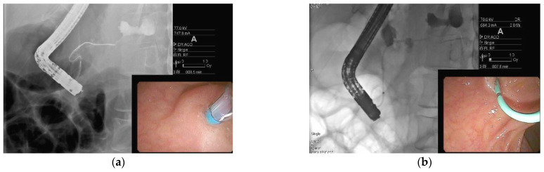
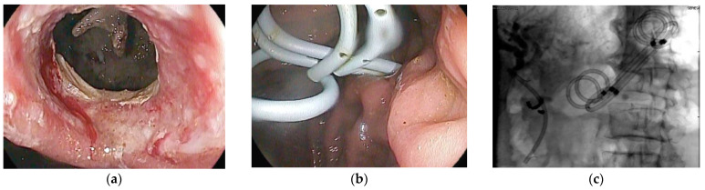
Pancreatic fluid collections (PFCs) are a common sequela of pancreatitis. Most PFCs can be managed conservatively, but symptomatic PFCs require either surgical, percutaneous, or endoscopic intervention. Recent advances in the therapeutics of PFCs, including the step-up approach, endoscopic ultrasound-guided transmural drainage with lumen apposing metal stents, and direct endoscopic necrosectomy, have ushered endoscopy to the forefront of PFCs management and have allowed for improved patient outcomes and decreased morbidity. In this review, we explore the progress and future of endoscopic management of PFCs.

Keywords: direct endoscopic necrosectomy; disconnected duct syndrome; dual-modality drainage; lumen-apposing metal stent; multiple transluminal gateway technique; necrotizing pancreatitis; pancreatic fluid collection; pseudocyst.


Conflict of interest statement

The authors declare no conflict of interest.

Figures

Figure 1
Figure 1
Endoscopic ultrasound image of a pseudocyst.
Figure 2
Figure 2
Computed tomography (CT) imaging of pancreatic pseudocyst. (a) Sagittal CT pancreas protocol image of a large 14.7 cm × 19.3 cm pseudocyst prior to lumen-apposing metal stent (LAMS) placement; (b) coronal CT image of pancreatic pseudocyst one day post-LAMS placement; and (c) coronal CT image of pancreatic pseudocyst six weeks post-LAMS placement.
Figure 3
Figure 3
CT and endoscopic image of a hemorrhagic pseudocyst. (a) Sagittal CT imaging of pseudocyst with heterogenous material consistent with hemorrhage; (b) coronal CT imaging of hemorrhagic pseudocyst prior to LAMS placement; and (c) endoscopic imaging post-LAMS placement with blood tinged fluid draining in the stomach.
Figure 4
Figure 4
Endoscopic ultrasound-guided drainage using a LAMS with subsequent direct endoscopic necrosectomy for walled-off pancreatic necrosis (WOPN) (a) Endoscopic ultrasound imaging of cyst cavity with necrotic debris; (b) endoscopic ultrasound-guided placement of LAMS into WOPN; (c) endoscopic imaging post LAMS deployment in the stomach; (d) endoscopic imaging inside cyst cavity with necrotic material; and (e) direct endoscopic necrosectomy performed using a snare.
Figure 5
Figure 5
Endoscopic retrograde pancreatography in a patient with disconnected duct syndrome (DDS). (a) Pancreatogram showing extravasation of contrast from the pancreatic duct in the body of the pancreas. The pancreatic duct in the tail is not opacified. The above findings are suggestive of complete DDS; and (b) pancreatic sphincterotomy followed by pancreatic duct stent placement.
Figure 6
Figure 6
Disconnected duct syndrome with LAMS removal and placement of double pigtail stents. (a) Endoscopic imaging showing LAMS removal with a residual portal into cyst cavity in a patient with known disconnected duct syndrome; (b) transmural plastic double-pigtail stents placed inside the cyst cavity; (c) fluoroscopic image showing double-pigtail stents to prevent recurrence of pancreatic fluid collection.

References

    1. Tyberg A., Karia K., Gabr M., Desai A., Doshi R., Gaidhane M., Sharaiha R.Z., Kahaleh M. Management of pancreatic fluid collections: A comprehensive review of the literature. World J. Gastroenterol. 2016;22:2256. doi: 10.3748/wjg.v22.i7.2256. - DOI - PMC - PubMed
    1. Li J., Yu Y., Li P., Zhang S.-T. Advancements in the endoscopic treatment of pancreatic fluid collections. Chronic Dis. Transl. Med. 2020;6:158–164. doi: 10.1016/j.cdtm.2020.03.001. - DOI - PMC - PubMed
    1. Bradley E.L., 3rd The natural and unnatural history of pancreatic fluid collections associated with acute pancreatitis. Dig. Dis. Sci. 2014;59:908–910. doi: 10.1007/s10620-013-3012-3. - DOI - PubMed
    1. Bradley E.L., 3rd, Clements J.L., Jr. Implications of diagnostic ultrasound in the surgical management of pancreatic pseudocysts. Am. J. Surg. 1974;127:163–173. doi: 10.1016/0002-9610(74)90153-6. - DOI - PubMed
    1. Bradley E.L. A clinically based classification system for acute pancreatitis: Summary of the International Symposium on Acute Pancreatitis, Atlanta, Ga, September 11 through 13, 1992. Arch. Surg. 1993;128:586–590. doi: 10.1001/archsurg.1993.01420170122019. - DOI - PubMed

LinkOut - more resources