

Gold Nanoparticles Functionalized with Angiogenin for Wound Care Application
- PMID: 33466813
- PMCID: PMC7830515
- DOI: 10.3390/nano11010201
Gold Nanoparticles Functionalized with Angiogenin for Wound Care Application
Abstract
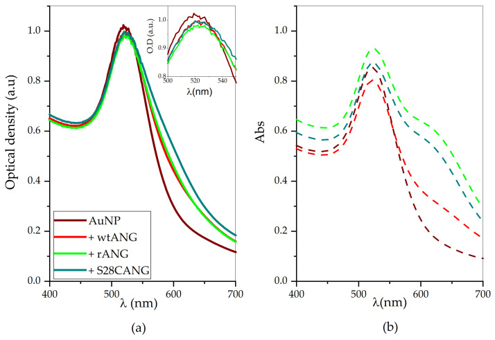
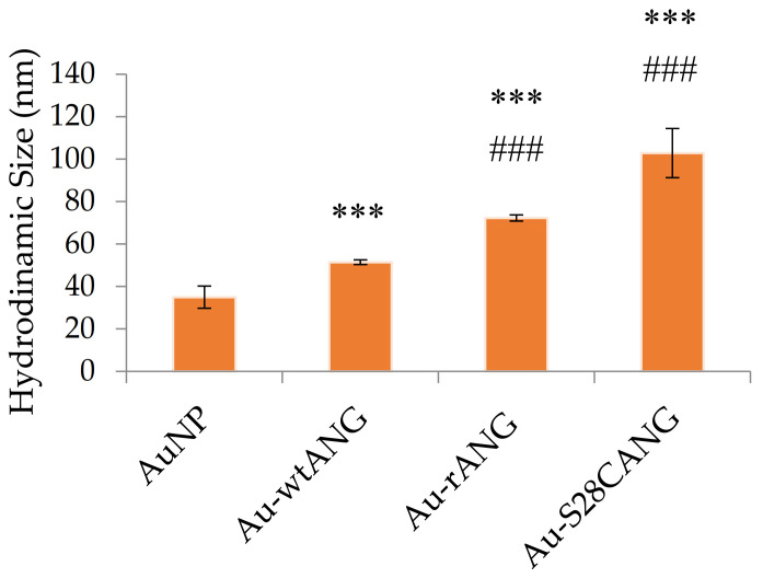
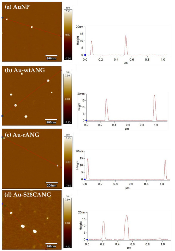
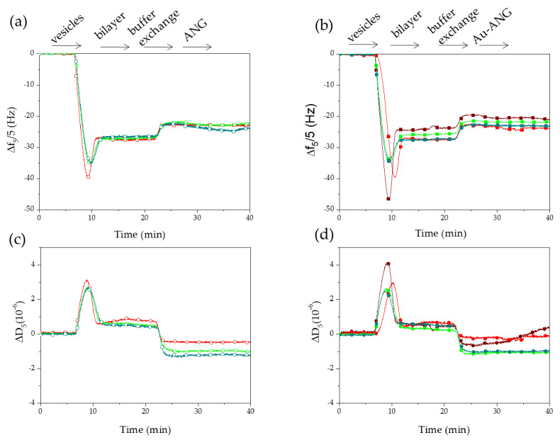
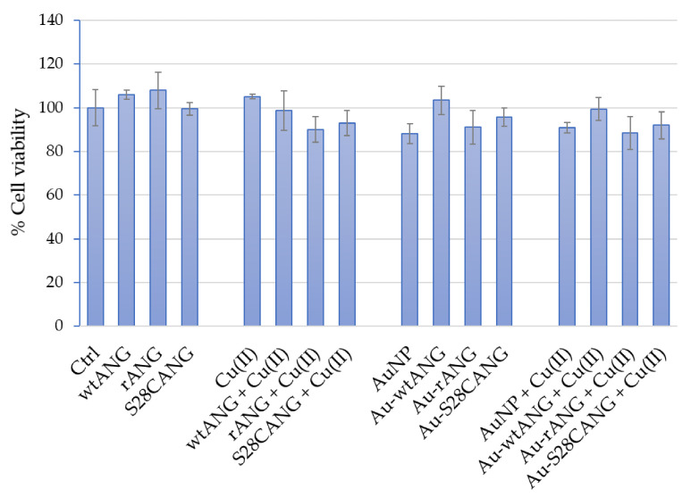
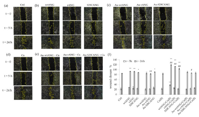
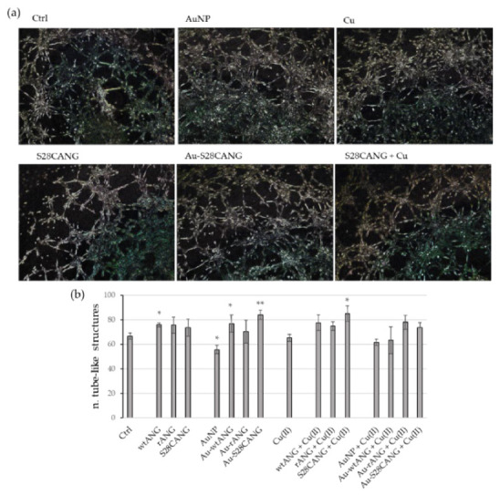
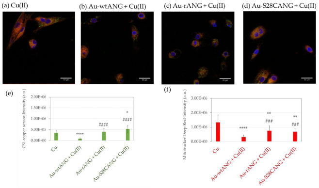
In this work, we aimed to develop a hybrid theranostic nano-formulation based on gold nanoparticles (AuNP)-having a known anti-angiogenic character-and the angiogenin (ANG), in order to tune the angiogenesis-related phases involved in the multifaceted process of the wound healing. To this purpose, spherical were surface "decorated" with three variants of the protein, namely, the recombinant (rANG), the wild-type, physiologically present in the human plasma (wtANG) and a new mutant with a cysteine substitution of the serine at the residue 28 (S28CANG). The hybrid biointerface between AuNP and ANG was scrutinized by a multi-technique approach based on dynamic light scattering, spectroscopic (UV-visible, circular dichroism) and microscopic (atomic force and laser scanning confocal) techniques. The analyses of optical features of plasmonic gold nanoparticles allowed for discrimination of different adsorption modes-i.e.; predominant physisorption and/or chemisorption-triggered by the ANG primary sequence. Biophysical experiments with supported lipid bilayers (SLB), an artificial model of cell membrane, were performed by means of quartz crystal microbalance with dissipation monitoring acoustic sensing technique. Cellular experiments on human umbilical vein endothelial cells (HUVEC), in the absence or presence of copper-another co-player of angiogenesis-were carried out to assay the nanotoxicity of the hybrid protein-gold nanoassemblies as well as their effect on cell migration and tubulogenesis. Results pointed to the promising potential of these nanoplatforms, especially the new hybrid Au-S28CANG obtained with the covalent grafting of the mutant on the gold surface, for the modulation of angiogenesis processes in wound care.
Keywords: AFM; QCM-D; angiogenesis; confocal microscopy; copper; endothelial cells; mutant protein; nanomaterial; nanomedicine; plasmonics.
Conflict of interest statement
The authors declare no conflict of interest.
Figures

Similar articles
-
Gold nanoparticles functionalized with angiogenin-mimicking peptides modulate cell membrane interactions.Biointerphases. 2018 Apr 16;13(3):03C401. doi: 10.1116/1.5022295. Biointerphases. 2018. PMID: 29660986
-
Graphene Oxide Nanosheets Tailored With Aromatic Dipeptide Nanoassemblies for a Tuneable Interaction With Cell Membranes.Front Bioeng Biotechnol. 2020 May 8;8:427. doi: 10.3389/fbioe.2020.00427. eCollection 2020. Front Bioeng Biotechnol. 2020. PMID: 32457892 Free PMC article.
-
Hyaluronan-Metal Gold Nanoparticle Hybrids for Targeted Tumor Cell Therapy.Int J Mol Sci. 2020 Apr 27;21(9):3085. doi: 10.3390/ijms21093085. Int J Mol Sci. 2020. PMID: 32349323 Free PMC article.
-
Probing the Interaction between Nanoparticles and Lipid Membranes by Quartz Crystal Microbalance with Dissipation Monitoring.Front Chem. 2016 Dec 5;4:46. doi: 10.3389/fchem.2016.00046. eCollection 2016. Front Chem. 2016. PMID: 27995125 Free PMC article. Review.
-
Lipid bilayers: Phase behavior and nanomechanics.Curr Top Membr. 2020;86:1-55. doi: 10.1016/bs.ctm.2020.08.005. Epub 2020 Sep 16. Curr Top Membr. 2020. PMID: 33837691 Review.
Cited by
-
Citrate-Coated Magnetic Polyethyleneimine Composites for Plasmid DNA Delivery into Glioblastoma.Polymers (Basel). 2021 Jul 6;13(14):2228. doi: 10.3390/polym13142228. Polymers (Basel). 2021. PMID: 34300986 Free PMC article.
-
Angiogenin and Copper Crossing in Wound Healing.Int J Mol Sci. 2021 Oct 2;22(19):10704. doi: 10.3390/ijms221910704. Int J Mol Sci. 2021. PMID: 34639045 Free PMC article. Review.
-
Pd-Based Hybrid Nanoparticles As Multimodal Theranostic Nanomedicine.ACS Appl Bio Mater. 2023 Feb 20;6(2):483-493. doi: 10.1021/acsabm.2c00759. Epub 2023 Jan 18. ACS Appl Bio Mater. 2023. PMID: 36651801 Free PMC article.
-
Peptides Derived from Angiogenin Regulate Cellular Copper Uptake.Int J Mol Sci. 2021 Sep 2;22(17):9530. doi: 10.3390/ijms22179530. Int J Mol Sci. 2021. PMID: 34502439 Free PMC article.
-
Effect of Citrate- and Gold-Stabilized Superparamagnetic Iron Oxide Nanoparticles on Head and Neck Tumor Cell Lines during Combination Therapy with Ionizing Radiation.Bioengineering (Basel). 2022 Dec 15;9(12):806. doi: 10.3390/bioengineering9120806. Bioengineering (Basel). 2022. PMID: 36551012 Free PMC article.
References
-
- Morimoto N., Kakudo N., Matsui M., Ogura T., Hara T., Suzuki K., Yamamoto M., Tabata Y., Kusumoto K. Exploratory clinical trial of combination wound therapy with a gelatin sheet and platelet-rich plasma in patients with chronic skin ulcers: Study protocol. BMJ Open. 2015;5:e007733. doi: 10.1136/bmjopen-2015-007733. - DOI - PMC - PubMed
-
- Houghton P.E. Electrical stimulation therapy to promote healing of chronic wounds: A review of reviews. Chronic Wound Care Manag. Res. 2017;4:25–44. doi: 10.2147/CWCMR.S101323. - DOI
Grants and funding
LinkOut - more resources
Full Text Sources
Other Literature Sources
Miscellaneous
