. 2021 Jan 14;22(2):802.
doi: 10.3390/ijms22020802.

Identification of the Primary Factors Determining theSpecificity of Human VKORC1 Recognition by Thioredoxin-Fold Proteins

Affiliations

Identification of the Primary Factors Determining theSpecificity of Human VKORC1 Recognition by Thioredoxin-Fold Proteins

Maxim Stolyarchuk et al. Int J Mol Sci. .

Abstract

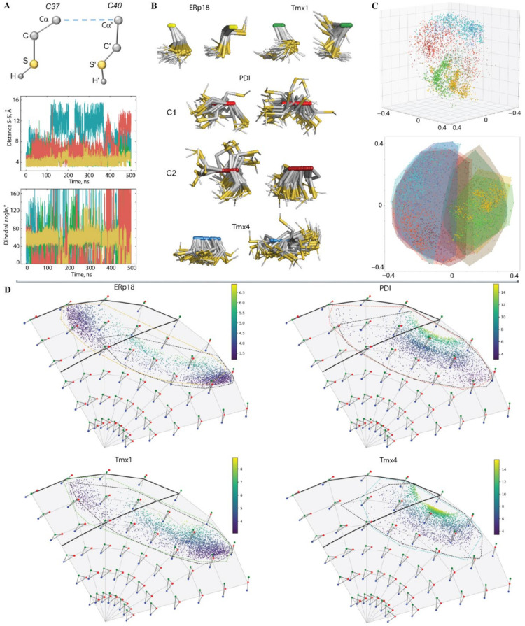
Redox (reduction-oxidation) reactions control many important biological processes in all organisms, both prokaryotes and eukaryotes. This reaction is usually accomplished by canonical disulphide-based pathways involving a donor enzyme that reduces the oxidised cysteine residues of a target protein, resulting in the cleavage of its disulphide bonds. Focusing on human vitamin K epoxide reductase (hVKORC1) as a target and on four redoxins (protein disulphide isomerase (PDI), endoplasmic reticulum oxidoreductase (ERp18), thioredoxin-related transmembrane protein 1 (Tmx1) and thioredoxin-related transmembrane protein 4 (Tmx4)) as the most probable reducers of VKORC1, a comparative in-silico analysis that concentrates on the similarity and divergence of redoxins in their sequence, secondary and tertiary structure, dynamics, intraprotein interactions and composition of the surface exposed to the target is provided. Similarly, hVKORC1 is analysed in its native state, where two pairs of cysteine residues are covalently linked, forming two disulphide bridges, as a target for Trx-fold proteins. Such analysis is used to derive the putative recognition/binding sites on each isolated protein, and PDI is suggested as the most probable hVKORC1 partner. By probing the alternative orientation of PDI with respect to hVKORC1, the functionally related noncovalent complex formed by hVKORC1 and PDI was found, which is proposed to be a first precursor to probe thiol-disulphide exchange reactions between PDI and hVKORC1.

Keywords: 3D modelling; PDI–hVKORC1 complex; Trx-fold proteins; dynamics; hVKORC1; molecular dynamics simulation; molecular recognition; protein folding; protein–protein interactions; thiol–disulphide exchange.


Conflict of interest statement

The authors declare no conflict of interest. The funders had no role in the design of the study; in the collection, analyses, or interpretation of data; in the writing of the manuscript, or in the decision to publish the results.

Figures

Figure 1
Figure 1
Thioredoxin-fold protein as a physiological reductant of human vitamin K epoxide reductase complex 1 (hVKORC1). (A) Oxidation of the two cysteine residues in the CX1X2C motif of Trx-fold proteins forms a disulphide bond, a process associated with the loss of two hydrogen atoms and, hence, two electrons (top). Mechanism of disulphide exchange between Trx and a target (bottom). The H-donor enzyme and a target are coloured in blue and green, respectively. (B) The Trx-fold is illustrated using the X-ray structure of human PDI deposed in PDB [5] (PDB ID: 4ekz). The protein is shown as red ribbons, with two cysteine residues from the CX1X2C motif as yellow balls. The four α-helices (in red), five β-strands (in yellow) and eight loops (in green) are numbered. (C) Comparison of the sequences of Trx-fold proteins ERp18, PDI, Tmx1 and Tmx4. Sequences were aligned on ERp18, having the most elongated sequence with ESPript3 (http://espript.ibcp.fr/). The solution with the best score is shown. The residues are coloured according to the consensus values: red indicates strict identity or similarity, while nonconserved residues are in black. Blue highlights the CX1X2C motif. (D) Ribbon diagram of the 3D human VKORC1 model in its inactive state showed in two orthogonal projections. The L-loop is shown in the colour teal, while disulphide bridges formed by cysteine residues C43—C51 and C132—C135 are drawn as yellow sticks. The transmembrane helices (TM) are numbered as in [6]. (E) The structure of VKOR from Synechococcus sp (bVKOR; ID PDB: 4nv5) is visualised using ribbons. The structural fragments that have sequences most similar to hVKORC1 and the Trx-like domain are shown in dark grey and light blue, respectively. The disulphide bridges formed by cysteine residues in Trx-like and VKOR-like domains are drawn as yellow sticks.
Figure 2
Figure 2
Characterisation of the MD simulations for the four Trx-fold proteins ERp18, PDI, Tmx1 and Tmx4. (A) RMSDs from the initial coordinates computed for all Cα-atoms (right) in each protein after fitting to initial conformation. (B) The superimposed average structures of each protein over replicas 1 and 2. Cysteine residues are shown as yellow balls. RMSD values of 0.5, 0.4, 0.3 and 0.4 Å in Erp18, PDI, Tmx1 and Tmx4, respectively. (C) RMSFs computed for the Cα-atoms using RMSF amplitude values less than 4 Å for the MD conformation of each protein after fitting to the initial conformation. Highly fluctuating residues (3, 6 and 5 in ERP18, Tmx1 and Tmx4, respectively) were excluded from the RMSD computation. In the insert, the secondary structures—αH- (red), 310-helices (light blue) and β-strands (dark blue)—were assigned for a mean conformation of every MD trajectory, 1 (top) and 2 (bottom), of each protein and were labelled as in the crystallographic structure of human PDI. (AC) Proteins are distinguished by colour (first/second replicas): ERp18 (yellow/brown), PDI (light/dark red), Tmx1 (light/dark green) and Tmx4 (light/dark blue). The numbering of the residues in each Trx-fold protein is arbitrary and starts from the first amino acid in the 3D model.
Figure 3
Figure 3
Intrinsic motion in the Trx-folded proteins and its interdependence. (A) Inter-residue cross-correlation maps computed for the Cα-atom pairs of ERp18, PDI, Tmx1 and Tmx4 after the fitting procedure. Secondary structure projected onto the protein sequences (α-helix/β-strand in red/blue) is shown at the border of matrices. Correlated (positive) and anticorrelated (negative) motions between Cα-atom pairs are shown as a red–blue gradient. (B) The PCA modes calculated for each protein after least-square fitting of the MD conformations to the average conformation as a reference. The bar chart gives the eigenvalue spectra in descending order for the first 10 modes (left). Projection of ERp18, PDI, Tmx1 and Tmx4 MD conformations with the principal component (PC) in 2D (middle) and 3D subspaces (right). MD conformations were taken every 100 ps (2D) and 10 ps (3D). The protein data is referenced by colour—ERp18 (dark yellow), PDI (brown), Tmx1 (green) and Tmx4 (dark blue and light blue for two replicas). (C) Collective motions characterised by the first two PCA modes. Atomic components in PCA modes 1–2 are drawn as red (1st mode) and cyan (2nd mode) arrows projected on a tube representation of each protein. For clarity, only motion with an amplitude ≥2 Å is represented. Cysteine residues are shown as yellow balls. All computations were performed on the Cα-atoms with RMSF fluctuations less than 4 Å for each protein after fitting on the initial conformation.
Figure 4
Figure 4
Sequence and folding of Trx-like proteins. (A) Alignment of the sequences and the secondary structure assigned to a mean conformation of the concatenated trajectory of each studied protein. Residues are coloured according to their properties—the positively and negatively charged residues are in red and blue, respectively; the hydrophobic residues are in green; the polar and amphipathic residues are in black; the CX1X2C motif is highlighted by a yellow background. The α-helices and β-strands are shown as red batons and yellow arrows, respectively. Secondary structure labelling is shown below the Tmx4 sequence. (B) The superimposed 3D structures of the Trx-fold proteins are shown in two orthogonal projections. The proteins are drawn as ribbons, with the cysteine residue as yellow balls. The F1 and F2 regions (and secondary structure labels) that are potentially involved in target recognition and/or the electron transfer reaction are outlined by dashed lines in (A) and differentiated by colour in (B) to distinguish between the proteins: ERp18 (dark yellow), PDI (red), Tmx1 (green) and Tmx4 (dark blue).
Figure 5
Figure 5
The CX1X2C motif geometries for ERp18, PDI, Tmx1 and Tmx4. (A) Geometry of the CX1X2C motif (left) is described by distance S⋯S’ (middle) and dihedral angle (right), determined as an absolute value of the pseudo torsion angle S−Cα(C37)−Cα’(C40)−S’. Only one replica 2 is shown. (B) Superposition of the thiol groups (Cα-C-S-H) from the CX1X2C motif of each protein is shown for either only one MD trajectory (ERP18, Tmx1 and Tmx4) or for both (PDI). Samples were taken for each 100-ns frame. (C) Multidimensional scaling (MDS) in 2D and 3D on the set of S-C-C-S tetrahedrons. Embedded points have been coloured according to the partner and replica they belong to. (D) Evolution of the shape of the triangles S-H⋯S on Kendall’s disk of 3D triangles; each data point is coloured according to the S⋯S distance. Representative triangles are regularly sampled on the disk. The thick black line delimits the area of conformations favouring H-bond interaction. The dashed areas are contouring subpopulations according to the S-atom being the H-donor.
Figure 6
Figure 6
hVKORC1 in its inactive state and its conventional MD simulations. (A) 3D model of hVKORC1 in its inactive state; it was inserted into the membrane (top) and zoomed in on the L-loop (bottom). The L-loop is highlighted by the colour teal; disulphide bridges formed by cysteine residues C43-C51 and C132-C135 are drawn as yellow sticks. Transmembrane helices (TMs) are numbered as in [6]. (B,C) RMSDs computed for each MD trajectory (replicas 1–3) from initial coordinates (at t = 0 ns, the same for all replicas) on the Cα-atoms of full-length hVKORC1 (in black, grey and rose brown), of the transmembrane domain (in orange, red and grenadine), of the L-loop (in clear aqua, bleu and navy) and of the N- and C-terminals (in teal, green and deep green) after fitting to the initial conformation of the respective fragment (B); of the L-loop (i) after fitting to its initial conformation (clear aqua, blue and navy blue) and (ii) after fitting of the protein coordinates to the initial conformation of the TMD (black, grey and silver) (C). (D) RMSFs computed for Cα-atoms of the MD conformations (replicas 13) after fitting to the initial conformation (at t = 0 ns, the same for all replicas; in black, grey and rose brown). In the insert, the folded secondary structures, αH- (red) and 310-helices (blue), were assigned for a mean conformation of each MD trajectory. (E) Superimposition of the L-loop conformations picked from replica 3 at 150 (grey), 250 (light blue) and 375 ns (deep teal). (F) The hVKORC1 sequence (Q9BQB6) and the secondary structure assignment for a mean conformation over each MD trajectory. Residues are coloured according to their properties: positively and negatively charged residues are in red and blue, respectively; hydrophobic residues are in green; polar and amphipathic residues are in black; residues C43, C51 and the CX1X2C motif are highlighted by a yellow background. α- and 310-helices are shown as red and blue batons, respectively. Secondary structure labelling is shown above the VKORC1 sequence. The L-loop sequence is surrounded by dashed lines.
Figure 7
Figure 7
Intrinsic motion of hVKORC1 and its L-loop. (A) The inter-residue cross-correlation map computed for the Cα-atom pairs after fitting to the respective first conformation (t = 0 ns) of the full-length hVKORC1 (top) and the L-loop (bottom) is shown for the three replicas. Correlated (positive) and anticorrelated (negative) motions between the Cα-atom pairs are shown as a red–blue gradient. (B) The PCA modes of the full-length hVKORC1 (top) and the L-loop (bottom), calculated for each MD trajectory after least-square fitting of the MD conformations to the average conformation of the respective domain as a reference. The bar plot gives the eigenvalue spectra in descending order for the first 10 modes. The data for replicas 13 are coloured black, grey and rose brown, respectively, while, for the full-length hVKORC1, the colouring is clear aqua, blue and navy blue for the L-loop. (C) Atomic components in the first PCA modes of the L-loop are drawn as red (1st mode) and blue (2nd mode) arrows projected onto the respective average structure from replicas 1 (top), 2 (middle) and 3 (bottom). Only motion with an amplitude ≥2Å is shown. The S-S bridge of hVKORC1 is shown using yellow sticks.
Figure 8
Figure 8
Geometry and folding of the L-loop from hVKORC1 in its inactive state. (A) Two tetrahedrons, T1—defined for the Cα-atom of C135 and for the midpoint residues of each L-loop helix, and T2—defined for the Cα-atom of C135 and for the most fluctuating residues (with the greatest RMSF values), from the L-loop linkers. (B) Distances between each pair of Cα-atoms from the tetrahedrons T1 (top) and T2 (bottom) over each MD trajectory. The distance curves and the edges of a tetrahedron are coloured similarly. (C) The time-dependent evolution of the secondary structure of each residue, as assigned by the Define Secondary Structure of Proteins (DSSP) method: α-helix is in red, 310-helix is in blue, turn is in orange and bend is in dark yellow. (D) Drift of the L-loop helices observed over the MD simulations (concatenated trajectory, sampled every 100 ps). Superimposed axes of helices from the L-loop are covered on the randomly chosen conformation of hVKORC1 in two orthogonal projections. The axis of each helix is defined as a line connecting the two centroids assigned for the first and the last residues.
Figure 9
Figure 9
Ensemble-based clustering of L-loop MD conformations. (A) Number of clusters obtained for each MD trajectory (1, 2 and 3) and the concatenated trajectory. The first 70 ns of every trajectory was omitted from the computation. Clustering was performed on each 10-ps frame of every trajectory using cut-off values that varied from 1.6 to 3.0 Å, with a step of 0.2 Å. (B) Location of the MD conformations grouped in clusters, with a cut-off of 2.0 Å for the RMSD curves of trajectories 13. Clusters C1–C6 are arbitrarily distinguished by colours in each trajectory: orange (C1), red (C2), blue (C3), rose (C4), green (C5) and violet (C6). (C) Representative conformations of the L-loop from clusters (Cm) with population ≥4%, obtained with a cut-off of 2.0 Å for the merged trajectory. The L-loop is shown as ribbons with a meshed surface, with disulphide bridges C43–C51 drawn as yellow sticks. The L-loop surface is displayed as meshed contours. The population of each cluster is given in brackets (in %), together with the replica number (in the bold) and the time (in ns) over which the representative conformation was recorded within a replica. (D) Conformations of the L-loop (taken every 100 frames) of each cluster (Cm) of the merged trajectory, and (E) superposed conformations from the C1m–C6m clusters. In (D,E), the L-loop is drawn as a tube.
Figure 10
Figure 10
Interacting residues in L-loop conformations. (A) Intraloop H-bond interactions in the L-loop conformations from clusters C1m–C6m. H-bonds D-H⋯A (D⋯A < 3.6 Å, ∠DHA ≥ 120°), where D and A are H-donor and H-acceptor (O/N) atoms, were analysed in a representative conformation from each cluster of the merged trajectories. Interactions that stabilised the helices were not considered. The L-loop is shown as ribbons, with the interacting residues as sticks and H-bond traces as dashed lines. Common H-bonding motifs are encircled by magenta (at R53), blue (at R61) and orange (at R37). The most characteristic donor and acceptor groups are labelled. N, O and C atoms are in blue, red and grey, respectively. (B) Charged and polar residues protruding from the L-loop. (C) Hydrophobic residues protruding from of the L-loop. The L-loop is shown as ribbons, with the residues exposed to the solvent displayed as sticks with a space-filling encounter. In (B,C), the N, O and C atoms are in blue, red and orange, respectively.
Figure 11
Figure 11
Modelling of human PDI–VKORC1 complex. (A) MD simulations of 3D model PDI–VKORC1 complexes were performed, with gradually diminished distance (from 12.5 to 8.0 Å) between the sulphur (S) atoms of C37 from PDI and of C43 from the L-loop of hVKORC1. PDI has two orientations with respect to VKORC1, with F1 (Model 1, left) and F2 (Model 2, right) positioned above the middle of the L-loop surface. Both models of the PDI–VKORC1 complex are shown as snapshots taken at t = 10, 60 and 80 ns, with different S⋯S distances. The reference residues and fragments are labelled. (B,C) Conformations of the PDI–VKORC1 complex, with two different PDI orientations, chosen at t = 10, 60 and 80 ns, and their superposition at all three times. In (AC), the proteins are depicted as ribbons or as ribbons and surfaces and are distinguished by colour: a red palette was used for PDI and a cyan palette for hVKORC1, both nuanced by the tonality from light to dark to distinguish the conformations chosen at t = 10, 60 and 80 ns.
Figure 12
Figure 12
Intermolecular contacts at the interface between PDI and hVKORC1 in two models of the PDI–hVKORC1 complex. The intermolecular H-bonds and hydrophobic contacts between PDI and VKORC1 in Model 1 (A, top) and Model 2 (B, top). (A,B) The proteins are shown as coloured ribbons: PDI in red and brown and VKORC1 in cyan (L-loop), with the interacting residues and thiol groups as sticks. The contacts are indicated by dashed lines: H-bonds in yellow and hydrophobic contacts in salmon. The structural fragments and residues participating in the contacts are labelled. Analysis of the intermolecular contacts was performed on conformations taken at t = 80 ns. (A,B) A pattern of H-bond (in blue) and hydrophobic (in orange) contacts between the PDI and hVKORC1 residues (bottom). Residues are coloured according to their properties: the positively and negatively charged residues are in red and blue, respectively; the hydrophobic residues are in green; the polar and amphipathic residues are in black.

Similar articles

Cited by

References

    1. Hatahet F., Ruddock L.W. Protein Disulfide Isomerase: A Critical Evaluation of Its Function in Disulfide Bond Formation. Antioxidants Redox Signal. 2009;11:2807–2850. doi: 10.1089/ars.2009.2466. - DOI - PubMed
    1. Lee S., Kim S.M., Lee R.T. Thioredoxin and Thioredoxin Target Proteins: From Molecular Mechanisms to Functional Significance. Antioxidants Redox Signal. 2013;18:1165–1207. doi: 10.1089/ars.2011.4322. - DOI - PMC - PubMed
    1. Hudson D.A., Gannon S.A., Thorpe C. Oxidative protein folding: From thiol–disulfide exchange reactions to the redox poise of the endoplasmic reticulum. Free Radic. Biol. Med. 2015;80:171–182. doi: 10.1016/j.freeradbiomed.2014.07.037. - DOI - PMC - PubMed
    1. Winther J.R., Thorpe C. Quantification of thiols and disulfides. Biochim. Biophys. Acta. 2014;1840:838–846. doi: 10.1016/j.bbagen.2013.03.031. - DOI - PMC - PubMed
    1. Berman H.M., Westbrook J., Feng Z., Gilliland G., Bhat T.N., Weissig H., Shindyalov I.N., Bourne P.E. The Protein Data Bank. Nucleic Acids Res. 2000;28:235–242. doi: 10.1093/nar/28.1.235. - DOI - PMC - PubMed

MeSH terms

LinkOut - more resources