

Domains and Functions of Spike Protein in Sars-Cov-2 in the Context of Vaccine Design
- PMID: 33466921
- PMCID: PMC7829931
- DOI: 10.3390/v13010109
Domains and Functions of Spike Protein in Sars-Cov-2 in the Context of Vaccine Design
Abstract
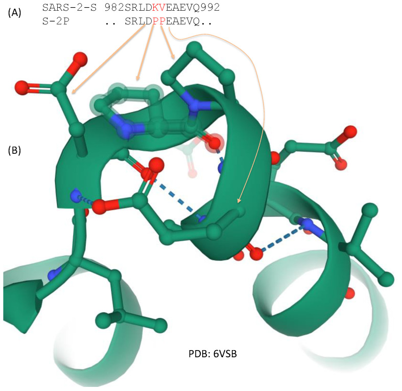
The spike protein in SARS-CoV-2 (SARS-2-S) interacts with the human ACE2 receptor to gain entry into a cell to initiate infection. Both Pfizer/BioNTech's BNT162b2 and Moderna's mRNA-1273 vaccine candidates are based on stabilized mRNA encoding prefusion SARS-2-S that can be produced after the mRNA is delivered into the human cell and translated. SARS-2-S is cleaved into S1 and S2 subunits, with S1 serving the function of receptor-binding and S2 serving the function of membrane fusion. Here, I dissect in detail the various domains of SARS-2-S and their functions discovered through a variety of different experimental and theoretical approaches to build a foundation for a comprehensive mechanistic understanding of how SARS-2-S works to achieve its function of mediating cell entry and subsequent cell-to-cell transmission. The integration of structure and function of SARS-2-S in this review should enhance our understanding of the dynamic processes involving receptor binding, multiple cleavage events, membrane fusion, viral entry, as well as the emergence of new viral variants. I highlighted the relevance of structural domains and dynamics to vaccine development, and discussed reasons for the spike protein to be frequently featured in the conspiracy theory claiming that SARS-CoV-2 is artificially created.
Keywords: COVID-19; S-2P; SARS-CoV-2; cleavage; hydrophobicity; isoelectric point; protein structure; spike protein; vaccine.
Conflict of interest statement
The author declares no conflict of interest.
Figures
Similar articles
-
Spike protein fusion loop controls SARS-CoV-2 fusogenicity and infectivity.J Struct Biol. 2021 Jun;213(2):107713. doi: 10.1016/j.jsb.2021.107713. Epub 2021 Mar 1. J Struct Biol. 2021. PMID: 33662570 Free PMC article.
-
Dynamics of SARS-CoV-2 Spike Proteins in Cell Entry: Control Elements in the Amino-Terminal Domains.mBio. 2021 Aug 31;12(4):e0159021. doi: 10.1128/mBio.01590-21. Epub 2021 Aug 3. mBio. 2021. PMID: 34340537 Free PMC article.
-
SARS-CoV-2 S protein:ACE2 interaction reveals novel allosteric targets.Elife. 2021 Feb 8;10:e63646. doi: 10.7554/eLife.63646. Elife. 2021. PMID: 33554856 Free PMC article.
-
Biological and Clinical Consequences of Integrin Binding via a Rogue RGD Motif in the SARS CoV-2 Spike Protein.Viruses. 2021 Jan 20;13(2):146. doi: 10.3390/v13020146. Viruses. 2021. PMID: 33498225 Free PMC article. Review.
-
The Spike of SARS-CoV-2: Uniqueness and Applications.Front Immunol. 2021 Jul 8;12:663912. doi: 10.3389/fimmu.2021.663912. eCollection 2021. Front Immunol. 2021. PMID: 34305894 Free PMC article. Review.
Cited by
-
Dramatic Differences between the Structural Susceptibility of the S1 Pre- and S2 Postfusion States of the SARS-CoV-2 Spike Protein to External Electric Fields Revealed by Molecular Dynamics Simulations.Viruses. 2023 Dec 11;15(12):2405. doi: 10.3390/v15122405. Viruses. 2023. PMID: 38140646 Free PMC article.
-
SARS-CoV-2 Proteins Interact with Alpha Synuclein and Induce Lewy Body-like Pathology In Vitro.Int J Mol Sci. 2022 Mar 21;23(6):3394. doi: 10.3390/ijms23063394. Int J Mol Sci. 2022. PMID: 35328814 Free PMC article.
-
Potency, toxicity and protection evaluation of PastoCoAd candidate vaccines: Novel preclinical mix and match rAd5 S, rAd5 RBD-N and SOBERANA dimeric-RBD protein.Vaccine. 2022 May 3;40(20):2856-2868. doi: 10.1016/j.vaccine.2022.03.066. Epub 2022 Apr 4. Vaccine. 2022. PMID: 35393148 Free PMC article.
-
An Immunological Review of SARS-CoV-2 Infection and Vaccine Serology: Innate and Adaptive Responses to mRNA, Adenovirus, Inactivated and Protein Subunit Vaccines.Vaccines (Basel). 2022 Dec 26;11(1):51. doi: 10.3390/vaccines11010051. Vaccines (Basel). 2022. PMID: 36679897 Free PMC article. Review.
-
An evolutionary insight into Severe Acute Respiratory Syndrome Coronavirus 2 Omicron variant of concern.Virus Res. 2022 Jun;314:198753. doi: 10.1016/j.virusres.2022.198753. Epub 2022 Mar 22. Virus Res. 2022. PMID: 35331836 Free PMC article.
References
Publication types
MeSH terms
Substances
Grants and funding
LinkOut - more resources
Full Text Sources
Other Literature Sources
Medical
Miscellaneous
