Review
. 2021 Jan 16;22(2):867.
doi: 10.3390/ijms22020867.

Possible Role of Metformin as an Immune Modulator in the Tumor Microenvironment of Ovarian Cancer

Affiliations
Review

Possible Role of Metformin as an Immune Modulator in the Tumor Microenvironment of Ovarian Cancer

Faye K Tsogas et al. Int J Mol Sci. .

Abstract

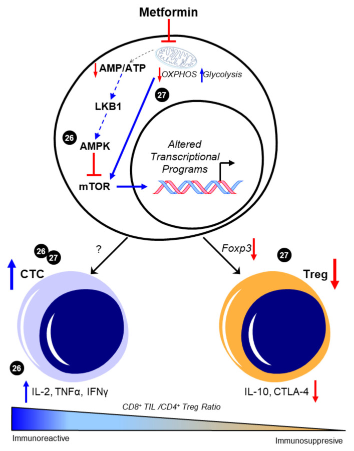
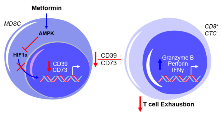
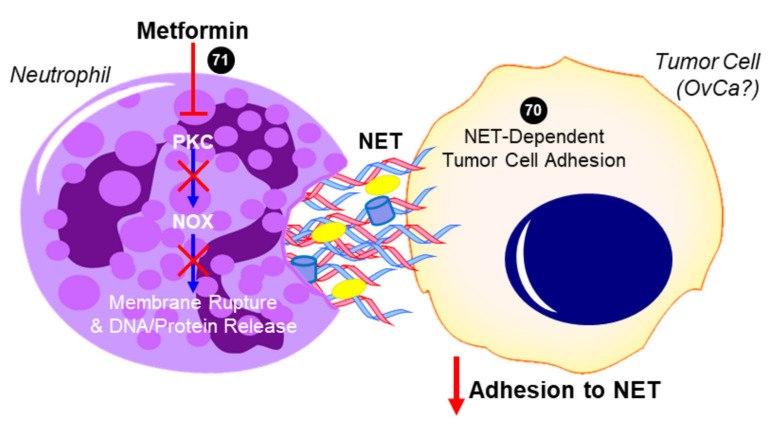
Growing evidence suggests that the immune component of the tumor microenvironment (TME) may be highly involved in the progression of high-grade serous ovarian cancer (HGSOC), as an immunosuppressive TME is associated with worse patient outcomes. Due to the poor prognosis of HGSOC, new therapeutic strategies targeting the TME may provide a potential path forward for preventing disease progression to improve patient survival. One such postulated approach is the repurposing of the type 2 diabetes medication, metformin, which has shown promise in reducing HGSOC tumor progression in retrospective epidemiological analyses and through numerous preclinical studies. Despite its potential utility in treating HGSOC, and that the immune TME is considered as a key factor in the disease's progression, little data has definitively shown the ability of metformin to target this component of the TME. In this brief review, we provide a summary of the current understanding of the effects of metformin on leukocyte function in ovarian cancer and, coupled with data from other related disease states, posit the potential mechanisms by which the drug may enhance the anti-tumorigenic effects of immune cells to improve HGSOC patient survival.

Keywords: T cell; macrophage; metformin; myeloid-derived suppressor cell; neutrophil; omentum; ovarian cancer; tumor microenvironment.


Conflict of interest statement

The authors declare that there are no competing interest.

Figures

Figure 1
Figure 1
Metformin enhances CD8+/CD4+ ratio to favor an immunoreactive microenvironment. The canonical pathway of metformin intracellular activity that results in 5′-AMP-activated protein kinase (AMPK)-dependent inhibition of the mammalian target of rapamycin (mTOR) is shown. Presumably, this enhancement in AMPK and the consequent mTOR inhibition is attributed to the metabolic effects of metformin in promoting glycolysis through inhibited mitochondrial respiration; however, this was not experimentally shown in these studies and is represented by dotted lines. The ability of metformin to increase the CD8+/CD4+ ratio [26,27] was associated with increases in functional CD8+ tumor-infiltrating lymphocytes (TILs) and concomitant increases in interleukin 2 (IL-2), tumor necrosis factor alpha (TNFα), and interferon gamma (IFNγ) [26]. Decreases in Foxp3-driven CD4+ T cell phenotypes and the subsequent reduction in interleukin 10 (IL-10) and cytotoxic T lymphocyte antigen-4 (CTLA-4) expression were also separately observed [27]. Cited experiments are denoted by their respective reference numbers [26,27]. CTC: cytotoxic CD8+ TILs, OXPHOS: oxidative phosphorylation, Treg: regulatory T cells. Blue arrows indicate upregulation/activation, red arrows indicate downregulation/inactivation and red T symbols indicate direct inhibition. Solid lines represent data presented in the cited manuscript; dotted lines indicate informed interactions from well-established data.
Figure 2
Figure 2
Metformin prevents myeloid-derived suppressor cell (MDSC)-mediated T cell exhaustion by downregulating CD39 and CD73 expression. The exposure of MDSCs to metformin activates AMPK and inhibits hypoxia-inducible factor 1-alpha (HIF1α) expression, thereby inhibiting the HIF1α-mediated transcriptional upregulation of CD39 and CD73 and subsequently suppressing cytotoxic CD8+ T lymphocytes (CTCs). This results in a net effect of reduced T cell exhaustion. Blue arrows indicate upregulation/activation, red arrows indicate downregulation/inactivation and red T symbols indicate inhibition.
Figure 3
Figure 3
Proposed role of metformin in inhibiting neutrophil extracellular trap (NET)-induced tumor cell metastasis. In addition to decreasing neutrophil recruitment (not shown), metformin has been shown to inhibit protein kinase C beta II (PKCβII) translocation to the plasma membrane and prevent the downstream activation of NADPH oxidase (NOX) that would otherwise result in the expulsion of neutrophil extracellular trap (NET) components into the extracellular space [71]. Tumor cells have been shown to adhere to NETs, which contain DNA, histones, neutrophil elastase, and other proteins. In the case of ovarian cancer (OvCa), NET formation was especially critical to the ability of tumor cells to colonize the omentum in vivo, while having no effect on the primary tumor or other intra-abdominal sites [70]. The ability for metformin to inhibit this NET-dependent adhesion and colonization of the omentum has not yet been investigated but is a potential mechanism for its antitumor activity. Cited experiments are denoted by their respective reference numbers [70,71]. Blue arrows indicate activation, red arrows indicate inhibition, red T symbols indicate direct inactivation, and red X symbols indicate inhibition of this pathway by metformin.

References

    1. Siegel R.L., Miller K.D., Jemal A. Cancer statistics, 2020. CA Cancer J. Clin. 2020;70:7–30. doi: 10.3322/caac.21590. - DOI - PubMed
    1. Doubeni C.A., Doubeni A.R., Myers A.E. Diagnosis and management of ovarian cancer. Am. Fam. Physician. 2016;93:937–944. - PubMed
    1. Surveillance, Epidemiology, and End Results (SEER) Program [(accessed on 10 November 2020)];Cancer Stat Facts: Ovarian Cancer. SEER* State Database: Mortality—All COD, Aggregated with State, Total, U.S. (1969–2017); Underlying Mortality Data Provided by NCHS. Available online: www.seer.cancer.gov/statfacts/html/ovary.html; www.cdc.gov/nchs.
    1. Winter W.E., Maxwell G.L., Tian C., Sundborg M.J., Rose G.S., Rose P.G., Rubin S.C., Muggia F.M., McGuire W.P. Tumor residual after surgical cytoreduction in prediction of clinical outcome in stage iv epithelial ovarian cancer: A gynecologic oncology group study. J. Clin. Oncol. 2008;26:83–89. doi: 10.1200/JCO.2007.13.1953. - DOI - PubMed
    1. Winter W.E., Maxwell G.L., Tian C., Carlson J.W., Ozols R.F., Rose P.G., Markman M., Armstrong D.K., Muggia F., McGuire W.P. Prognostic factors for stage iii epithelial ovarian cancer: A gynecologic oncology group study. J. Clin. Oncol. 2007;25:3621–3627. doi: 10.1200/JCO.2006.10.2517. - DOI - PubMed

LinkOut - more resources