

Maternal Folic Acid Deficiency Is Associated to Developing Nasal and Palate Malformations in Mice
- PMID: 33467180
- PMCID: PMC7830789
- DOI: 10.3390/nu13010251
Maternal Folic Acid Deficiency Is Associated to Developing Nasal and Palate Malformations in Mice
Abstract
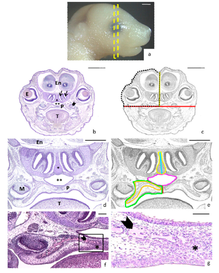
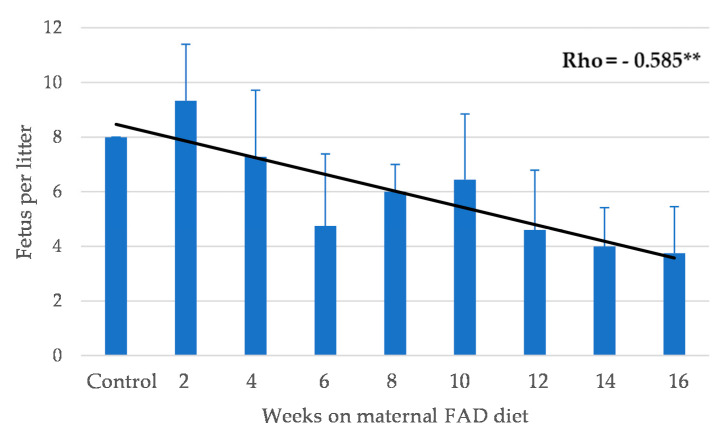
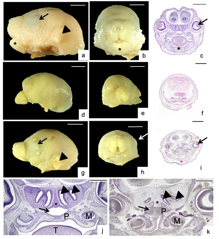
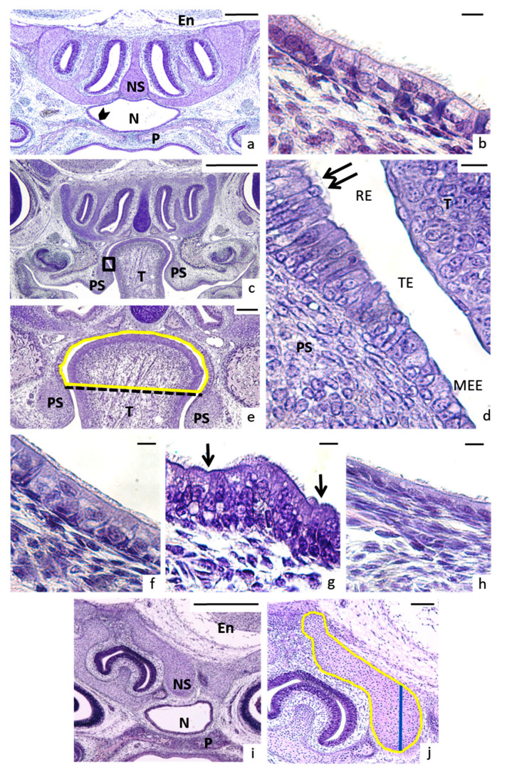
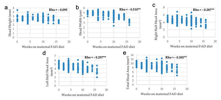
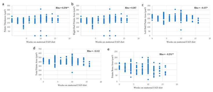
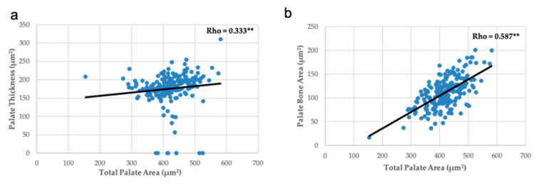
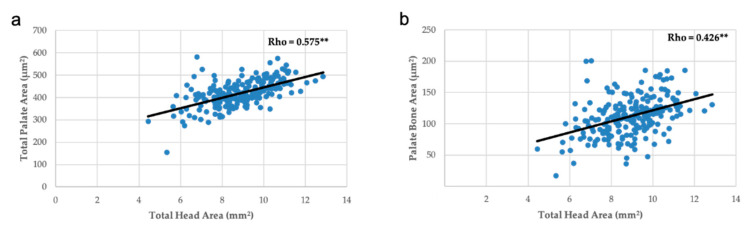
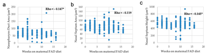
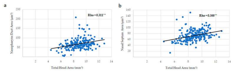
Craniofacial development requires extremely fine-tuned developmental coordination of multiple specialized tissues. It has been evidenced that a folate deficiency (vitamin B9), or its synthetic form, folic acid (FA), in maternal diet could trigger multiple craniofacial malformations as oral clefts, tongue, or mandible abnormalities. In this study, a folic acid-deficient (FAD) diet was administered to eight-week-old C57/BL/6J female mouse for 2-16 weeks. The head symmetry, palate and nasal region were studied in 24 control and 260 experimental fetuses. Our results showed a significant reduction in the mean number of fetuses per litter according to maternal weeks on FAD diet (p < 0.01). Fetuses were affected by cleft palate (3.8%) as well as other severe congenital abnormalities, for the first time related to maternal FAD diet, as head asymmetries (4.6%), high arched palate (3.5%), nasal septum malformed (7.3%), nasopharynx duct shape (15%), and cilia and epithelium abnormalities (11.2% and 5.8%). Dysmorphologies of the nasal region were the most frequent, appearing at just four weeks following a maternal FAD diet. This is the first time that nasal region development is experimentally related to this vitamin deficiency. In conclusion, our report offers novel discoveries about the importance of maternal folate intake on midface craniofacial development of the embryos. Moreover, the longer the deficit lasts, the more serious the consequent effects appear to be.
Keywords: congenital abnormalities; maternal folic acid-deficient diet; nasal region; palate.
Conflict of interest statement
The authors declare no conflict of interest.
Figures
Similar articles
-
Craniofacial structure alterations of foetuses from folic acid deficient pregnant mice.Ann Anat. 2018 Jul;218:59-68. doi: 10.1016/j.aanat.2018.02.010. Epub 2018 Mar 28. Ann Anat. 2018. PMID: 29604388
-
Tongue Abnormalities Are Associated to a Maternal Folic Acid Deficient Diet in Mice.Nutrients. 2017 Dec 28;10(1):26. doi: 10.3390/nu10010026. Nutrients. 2017. PMID: 29283374 Free PMC article.
-
Occurrence of cleft-palate and alteration of Tgf-β(3) expression and the mechanisms leading to palatal fusion in mice following dietary folic-acid deficiency.Cells Tissues Organs. 2011;194(5):406-20. doi: 10.1159/000323213. Epub 2011 Feb 2. Cells Tissues Organs. 2011. PMID: 21293104
-
Effects of folate and vitamin B12 deficiencies during pregnancy on fetal, infant, and child development.Food Nutr Bull. 2008 Jun;29(2 Suppl):S101-11; discussion S112-5. doi: 10.1177/15648265080292S114. Food Nutr Bull. 2008. PMID: 18709885 Review.
-
[Environment and genetics in the etiology of cleft lip and cleft palate with reference to the role of folic acid].Epidemiol Prev. 2000 Jan-Feb;24(1):21-7. Epidemiol Prev. 2000. PMID: 10748547 Review. Italian.
Cited by
-
Clinical Approach to Cleft Lip and Palate with or Without Surgical Correction in Ten Brachycephalic Puppies.Vet Sci. 2025 May 14;12(5):474. doi: 10.3390/vetsci12050474. Vet Sci. 2025. PMID: 40431567 Free PMC article.
-
A head start: The relationship of placental factors to craniofacial and brain development.Dev Dyn. 2025 Mar 19:10.1002/dvdy.70018. doi: 10.1002/dvdy.70018. Online ahead of print. Dev Dyn. 2025. PMID: 40105397 Review.
-
Relationship between maternal folic acid supplementation during pregnancy and risk of childhood asthma: Systematic review and dose-response meta-analysis.Front Pediatr. 2022 Nov 17;10:1000532. doi: 10.3389/fped.2022.1000532. eCollection 2022. Front Pediatr. 2022. PMID: 36467483 Free PMC article. Review.
References
-
- Sperber G.H. Craniofacial Development. BC Decker; Hamilton, ON, Canada: 2000.
-
- Mossey P., Castilla E. Report of a WHO Registry Meeting on Craniofacial Anomalies. World Health Organization; Geneva, Switzerland: 2003. Global Registry and Database on Craniofacial Anomalies.
MeSH terms
Grants and funding
LinkOut - more resources
Full Text Sources
Other Literature Sources
Medical
