Human Dental Pulp Tissue during Orthodontic Tooth Movement: An Immunofluorescence Study
- PMID: 33467280
- PMCID: PMC7739291
- DOI: 10.3390/jfmk5030065
Human Dental Pulp Tissue during Orthodontic Tooth Movement: An Immunofluorescence Study
Abstract
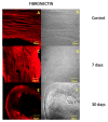
The orthodontic tooth movement is the last step of several biological processes that take place after the application of external forces. During this process, dental pulp tissue is subjected to structural and protein expression modifications in order to maintain their integrity and functional morphology. The purpose of the present work was to perform an in vivo study, evaluating protein expression modifications in the human dental pulp of patients that have undergone orthodontic tooth movement due to pre-calibrated light force application for 30 days. Dental pulp samples were extracted from molars and premolars of the control group and after 7 and 30 days of treatment; the samples were then processed for immunofluorescence reactions using antibodies against fibronectin, collagen I and vascular endothelial growth factor (VEGF). Our results show that, after 7 days of treatment, all tested proteins change their pattern expression and will reset after 30 days. These data demonstrate that the dental pulp does not involve any irreversible iatrogenic alterations, supporting the efficacy and safety of using pre-calibrated force application to induce orthodontic tooth movement in clinical practice.
Keywords: extracellular matrix proteins; human dental pulp; immunofluorescence; orthodontic tooth movement.
Conflict of interest statement
The Authors declare no conflict of interest.
Figures


References
-
- Anastasi G., Cordasco G., Matarese G., Nucera R., Rizzo G., Mazza M., Militi A., Portelli M., Cutroneo G., Favaloro A. An immunohistochemical, histological, and electron-microscopic study of the human periodontal ligament during orthodontic treatment. Int. J. Mol. Med. 2008;21:545–554. doi: 10.3892/ijmm.21.5.545. - DOI - PubMed
-
- Militi A., Cutroneo G., Favaloro A., Matarese G., Di Mauro D., Lauritano F., Centofanti A., Cervino G., Nicita F., Bramanti A., et al. An Immunofluorescence Study on VEGF and Extracellular Matrix Proteins in Human Periodontal Ligament during Tooth Movement. Heliyon. 2019;5:e02572. doi: 10.1016/j.heliyon.2019.e02572. - DOI - PMC - PubMed
-
- Cutroneo G., Centofanti A., Speciale F., Rizzo G., Favaloro A., Santoro G., Bruschetta D., Milardi D., Micali A., Di Mauro D., et al. Sarcoglycan Complex in Masseter and Sternocleidomastoid Muscles of Baboons: An Immunohistochemical Study. Eur. J. Histochem. 2015;59:2509. doi: 10.4081/ejh.2015.2509. - DOI - PMC - PubMed
-
- Cutroneo G., Vermiglio G., Centofanti A., Rizzo G., Runci M., Favaloro A., Piancino M.G., Bracco P., Ramieri G., Bianchi F., et al. Morphofunctional Compensation of Masseter Muscles in Unilateral Posterior Crossbite Patients. Eur. J. Histochem. 2016;60:2605. doi: 10.4081/ejh.2016.2605. - DOI - PMC - PubMed
LinkOut - more resources
Full Text Sources

