. 2021 Jan 15;10(1):156.
doi: 10.3390/cells10010156.

MRGPRX2 Activation by Rocuronium: Insights from Studies with Human Skin Mast Cells and Missense Variants

Affiliations

MRGPRX2 Activation by Rocuronium: Insights from Studies with Human Skin Mast Cells and Missense Variants

Chalatip Chompunud Na Ayudhya et al. Cells. .

Abstract

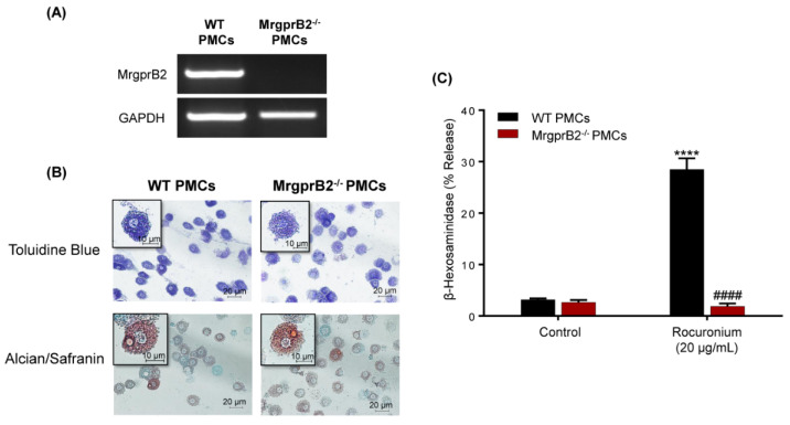
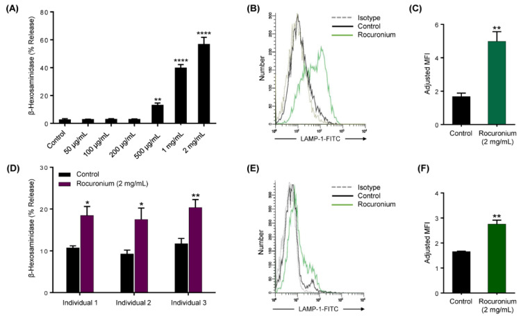
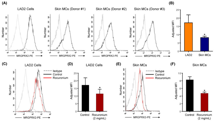
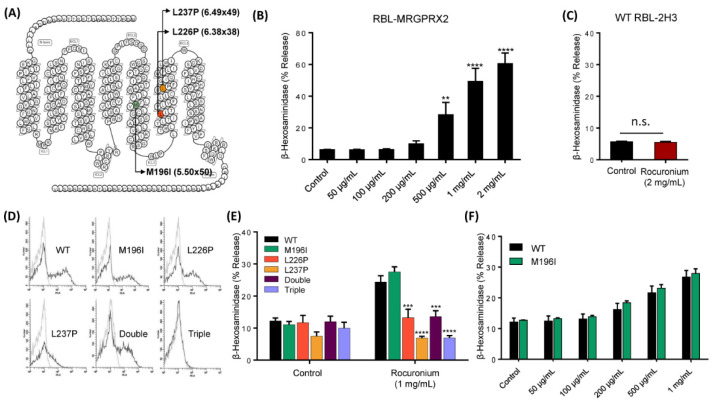
Perioperative hypersensitivity (POH) to the neuromuscular blocking drug (NMBD) rocuronium was previously thought to be IgE and mast cell (MC)-mediated. However, the recent seminal observation that rocuronium induces degranulation in murine peritoneal MCs (PMCs) via Mas-related G protein-coupled receptor B2 (MrgprB2) led to the idea that POH to this drug involves the activation of MRGPRX2 (human ortholog of MrgprB2). Furthermore, based on the demonstration that a patient with POH to rocuronium displayed three missense mutations (M196I, L226P and L237P) in MRGPRX2's transmembrane domains, it was proposed that this hypersensitivity reaction resulted from aberrant activation of this receptor. We found that rocuronium at 20 µg/mL caused degranulation in mouse PMCs via MrgprB2 but required at least 500 µg/mL to induce degranulation in human MCs via MRGPRX2. Furthermore, RBL-2H3 cells transiently expressing M196I, L226P and L237P variants did not display enhanced degranulation in response to rocuronium when compared to the wild-type receptor. These findings provide the first demonstration that rocuronium induces degranulation in human MCs via MRGPRX2. Furthermore, the important differences between MrgprB2 and MRGPRX2 and the inability of rocuronium to induce enhanced response in cells expressing MRGPRX2 variants suggest that the mechanism of its POH is more complex than previously thought.

Keywords: MRGPRX2; MrgprB2; anaphylaxis; mast cells; missense mutation; rocuronium.


Conflict of interest statement

The authors declare no conflict of interest.

Figures

Figure 1
Figure 1
Rocuronium activates mouse peritoneal mast cells (PMCs) via MrgprB2. (A) PCR analysis was performed to determine the mRNA expression of MrgprB2 in wild-type (WT) and MrgprB2−/− PMCs. GAPDH was used for normalization. (B) WT and MrgprB2−/− PMCs were stained with Toluidine blue and Alcian/Safranin. The images were acquired at 40× resolution. Bars = 10 and 20 μm. (C) WT and MrgprB2−/− PMCs were stimulated with rocuronium (20 μg/mL) for 30 min, and β-hexosaminidase release was determined. Data are expressed as mean ± SEM. Statistical significance was determined by two-way ANOVA with Tukey post-hoc test. **** p < 0.0001 compared to the control, #### p < 0.0001 compared between WT and MrgprB2−/− groups.
Figure 2
Figure 2
Rocuronium activates LAD2 cells and primary human skin mast cells (MCs) to cause degranulation. (A) LAD2 cells were exposed to buffer (control) or different concentrations of rocuronium for 30 min, and β-hexosaminidase release was determined. (B) Cells were stimulated with rocuronium (2 mg/mL) for 5 min, and LAMP-1 expression was determined by flow cytometry. Representative histograms of three independent experiments are shown. (C) The adjusted mean fluorescent intensity (MFI) levels of LAMP-1 expression are shown. Adjusted MFI was calculated as MFI of sample/MFI of isotype control. (D) Primary skin MCs were isolated and cultured from the human skin of 3 different donors. Skin-derived MCs were used to determine rocuronium-induced β-hexosaminidase release, and (E,F) LAMP-1 expression. All data points are the mean ± SEM of at least three experiments. For comparisons of two samples, a two-tailed unpaired t-test was used. For comparisons of multiple samples to a control group, one-way ANOVA with Dunnett’s post-hoc test was used. * p < 0.05, ** p < 0.01 and **** p < 0.0001.
Figure 3
Figure 3
Rocuronium induces MRGPRX2 internalization in LAD2 cells and primary human skin MCs. (A) Flow cytometry histograms for MRGPRX2 cell surface expression (solid black line) and isotype (dotted gray line) are shown. Histograms are representative of three independent experiments. (B) The comparison of MRGPRX2 expression levels between human MC line LAD2 cells and primary human skin MCs. (C) LAD2 cells were stimulated with rocuronium (2 mg/mL) for 30 min, and MRGPRX2 internalization was determined by flow cytometry. (D) The adjusted MFI levels of MRGPRX2 cell surface expression are shown. (E,F) Rocuronium-induced MRGPRX2 internalization and adjusted MFI levels of MRGPRX2 cell surface expression were determined in primary human skin MCs. All data points are the mean ± SEM of at least three experiments. Statistical significance was determined by two-tailed unpaired t-test. * p < 0.05.
Figure 4
Figure 4
MRGPRX2 mutations rendered the receptor unresponsiveness to rocuronium. (A) Snake diagram of MRGPRX2 indicating three missense mutations identified in the patient. (B) RBL-MRGPRX2 were stimulated with different concentrations of rocuronium for 30 min, and β-hexosaminidase release was determined. (C) Untransfected WT RBL-2H3 cells were stimulated with rocuronium (2 mg/mL) for 30 min, and β-hexosaminidase release was determined. (D) Cell surface expression of WT MRGPRX2 and its variants was determined by flow cytometry. (E) Cells expressing WT MRGPRX2 and its variants were exposed to buffer (control) or rocuronium (1 mg/mL) for 30 min, and β-hexosaminidase release was determined. (F) Dose response of rocuronium-induced β-hexosaminidase release was determined in cells expressing WT MRGPRX2 and M196I. Data are the mean ± SEM of at least three experiments. For comparisons of two samples, two-tailed unpaired t-test was used. For comparisons of multiple samples to a control group, one-way ANOVA with Dunnett’s post-hoc test was used. ** p < 0.01, *** p < 0.001 and **** p < 0.0001.

Similar articles

Cited by

References

    1. Volcheck G.W., Hepner D.L. Identification and Management of Perioperative Anaphylaxis. J. Allergy Clin. Immunol. Pract. 2019;7:2134–2142. doi: 10.1016/j.jaip.2019.05.033. - DOI - PubMed
    1. Van Gasse A.L., Elst J., Bridts C.H., Mertens C., Faber M., Hagendorens M.M., De Clerck L.S., Sabato V., Ebo D.G. Rocuronium Hypersensitivity: Does Off-Target Occupation of the MRGPRX2 Receptor Play a Role? J. Allergy Clin. Immunol. Pract. 2019;7:998–1003. doi: 10.1016/j.jaip.2018.09.034. - DOI - PubMed
    1. McNeil B.D., Pundir P., Meeker S., Han L., Undem B.J., Kulka M., Dong X. Identification of a mast-cell-specific receptor crucial for pseudo-allergic drug reactions. Nature. 2015;519:237–241. doi: 10.1038/nature14022. - DOI - PMC - PubMed
    1. Spoerl D., D’Incau S., Roux-Lombard P., Harr T., Czarnetzki C. Non-IgE-Dependent Hypersensitivity to Rocuronium Reversed by Sugammadex: Report of Three Cases and Hypothesis on the Underlying Mechanism. Int. Arch. Allergy Immunol. 2016;169:256–262. doi: 10.1159/000446182. - DOI - PubMed
    1. Suzuki Y., Liu S., Kadoya F., Takasaki Y., Yorozuya T., Mogi M. Association between mutated Mas-related G protein-coupled receptor-X2 and rocuronium-induced intraoperative anaphylaxis. Br. J. Anaesth. 2020 doi: 10.1016/j.bja.2020.05.046. - DOI - PubMed

Publication types

MeSH terms

LinkOut - more resources