Review
. 2021 Jan 15;10(1):161.
doi: 10.3390/cells10010161.

PE_PGRS33, an Important Virulence Factor of Mycobacterium tuberculosis and Potential Target of Host Humoral Immune Response

Affiliations
Review

PE_PGRS33, an Important Virulence Factor of Mycobacterium tuberculosis and Potential Target of Host Humoral Immune Response

Eliza Kramarska et al. Cells. .

Abstract

PE_PGRS proteins are surface antigens of Mycobacterium tuberculosis (Mtb) and a few other pathogenic mycobacteria. The PE_PGRS33 protein is among the most studied PE_PGRSs. It is known that the PE domain of PE_PGRS33 is required for the protein translocation through the mycobacterial cell wall, where the PGRS domain remains available for interaction with host receptors. Interaction with Toll like receptor 2 (TLR2) promotes secretion of inflammatory chemokines and cytokines, which are key in the immunopathogenesis of tuberculosis (TB). In this review, we briefly address some key challenges in the development of a TB vaccine and attempt to provide a rationale for the development of new vaccines aimed at fostering a humoral response against Mtb. Using PE_PGRS33 as a model for a surface-exposed antigen, we exploit the availability of current structural data using homology modeling to gather insights on the PGRS domain features. Our study suggests that the PGRS domain of PE_PGRS33 exposes four PGII sandwiches on the outer surface, which, we propose, are directly involved through their loops in the interactions with the host receptors and, as such, are promising targets for a vaccination strategy aimed at inducing a humoral response.

Keywords: infectious disease; protein structure; tuberculosis; vaccine.


Conflict of interest statement

The authors declare no conflict of interest. The funders had no role in the design of the study; in the collection, analyses, or interpretation of data; in the writing of the manuscript, or in the decision to publish the results.

Figures

Figure 1
Figure 1
Domain organization and sequence of PE_PGRS33. In the PE domain, the conserved PE motif is colored red. In the PGRS domain, sequences of the four PGII sandwich motifs are colored green, purple, blue, and cyan.
Figure 2
Figure 2
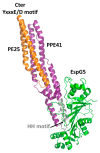
Cartoon representation of the crystal structure of PE25-PPE41 (orange/purple) ternary complex with the chaperone EspG5 (green). The signature ESX secretory motif YxxxE/D is located at the C-terminal side of PE25 (orange), whereas the EspG5 binding region is located on the HH motif of PPE41 (gray) [83].
Figure 3
Figure 3
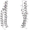
Cartoon representations of the homology model of the PE domain of PE_PGRS33. Front and side views are reported on left and right sides, respectively. The model was computed with MODELLER using the structure of the PE25 domain from a type VII secretion system of Mycobacterium tuberculosis (Mtb) as a template (PDB core 4w4k, sequence identity 37%). Hydrophobic and aromatic residues are drawn in stick representation.
Figure 4
Figure 4
Domain architecture of PE- or PPE-containing Mtb proteins (strain H37Rv), which are predicted to embed a serine hydrolase domain [93]. The last hydrolase, PE_PGRS35, is the Mtb homolog of M. marinum PecA [92].
Figure 5
Figure 5
Stick representation of the homology model of the PGII sandwich domain PGII4, computed using the structure of sfAFP as a template (pdb code 2pne). The front view (A) shows the six PGII helices, whereas the side view (B) shows the localization of hydrophobic residues (e.g., L432, I433, L461) on the lateral loops. The inset shows glycine residues pointing inside and stabilizing the tightly packed PGII helices.
Figure 6
Figure 6
Surface and stick representations of PGII domains in (A) PE_PGRS33 (domain PGII4); (B) antifreeze protein sfAFP (pdb code 2pne) in two 180° views; and (C) Salmonella bacteriophage S16 long tail fiber (pdb code 6F45). In this panel, the PGII domain is located at the C-terminal side of the protein (stick and surface representation). Adjacent domains are drawn in surface and cartoon representations. In all panels, the color code used for PGII residues is red for negative, blue for positive, green for hydrophobic, and light blue for polar residues.
Figure 7
Figure 7
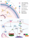
Schematic representations of (A) the PE_PGRS33 path to the mycobacterial membrane and (B) immune responses to PE_PGRS33 by the host.

Similar articles

Cited by

References

    1. Harding E. WHO global progress report on tuberculosis elimination. Lancet Respir. Med. 2020;8:19. doi: 10.1016/S2213-2600(19)30418-7. - DOI - PubMed
    1. World Health Organization The End TB Strategy. [(accessed on 14 January 2021)]; Available online: https://www.who.int/tb/strategy/en/
    1. Barry C.E., 3rd, Boshoff H.I., Dartois V., Dick T., Ehrt S., Flynn J., Schnappinger D., Wilkinson R.J., Young D. The spectrum of latent tuberculosis: Rethinking the biology and intervention strategies. Nat. Rev. Microbiol. 2009;7:845–855. doi: 10.1038/nrmicro2236. - DOI - PMC - PubMed
    1. Delogu G., Sali M., Fadda G. The biology of mycobacterium tuberculosis infection. Mediterr. J. Hematol. Infect. Dis. 2013;5:e2013070. doi: 10.4084/mjhid.2013.070. - DOI - PMC - PubMed
    1. Nikitushkin V.D., Demina G.R., Shleeva M.O., Guryanova S.V., Ruggiero A., Berisio R., Kaprelyants A.S. A product of RpfB and RipA joint enzymatic action promotes the resuscitation of dormant mycobacteria. Febs. J. 2015;282:2500–2511. doi: 10.1111/febs.13292. - DOI - PubMed

Publication types

MeSH terms

LinkOut - more resources