. 2021 Jan 15;13(2):270.
doi: 10.3390/polym13020270.

Additive-Manufactured Gyroid Scaffolds of Magnesium Oxide, Phosphate Glass Fiber and Polylactic Acid Composite for Bone Tissue Engineering

Affiliations

Additive-Manufactured Gyroid Scaffolds of Magnesium Oxide, Phosphate Glass Fiber and Polylactic Acid Composite for Bone Tissue Engineering

Lizhe He et al. Polymers (Basel). .

Abstract

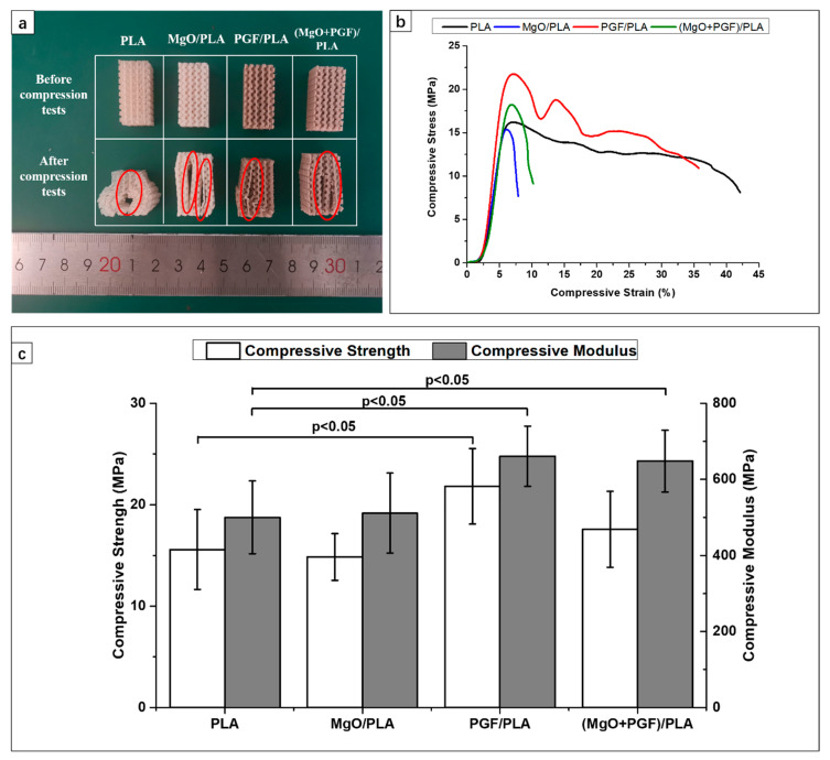
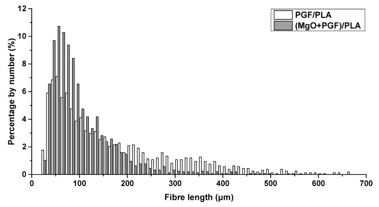
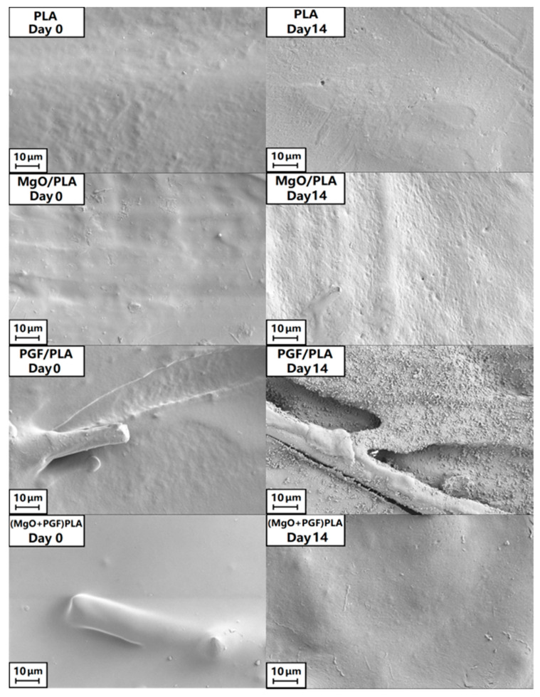
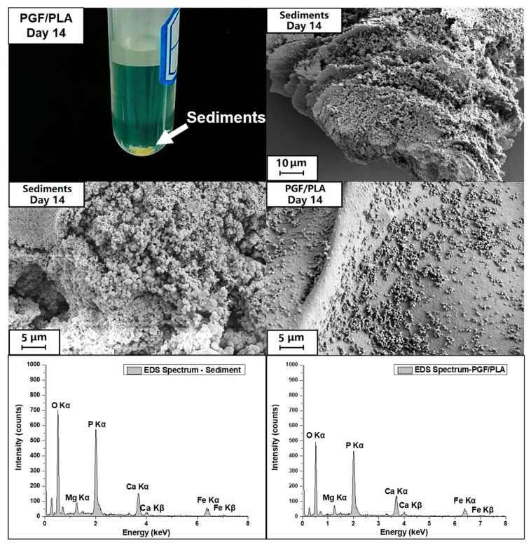
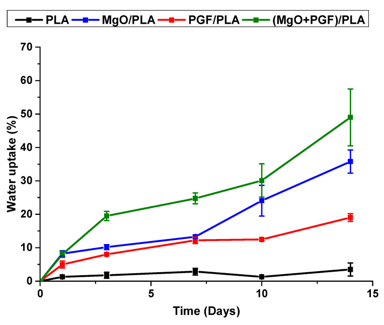
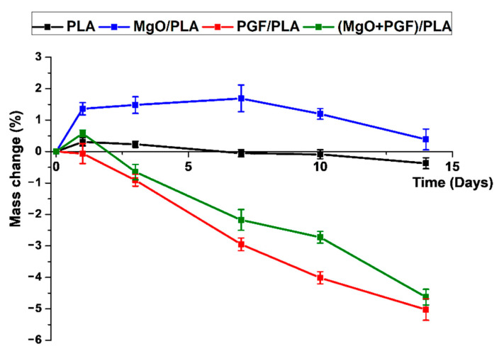
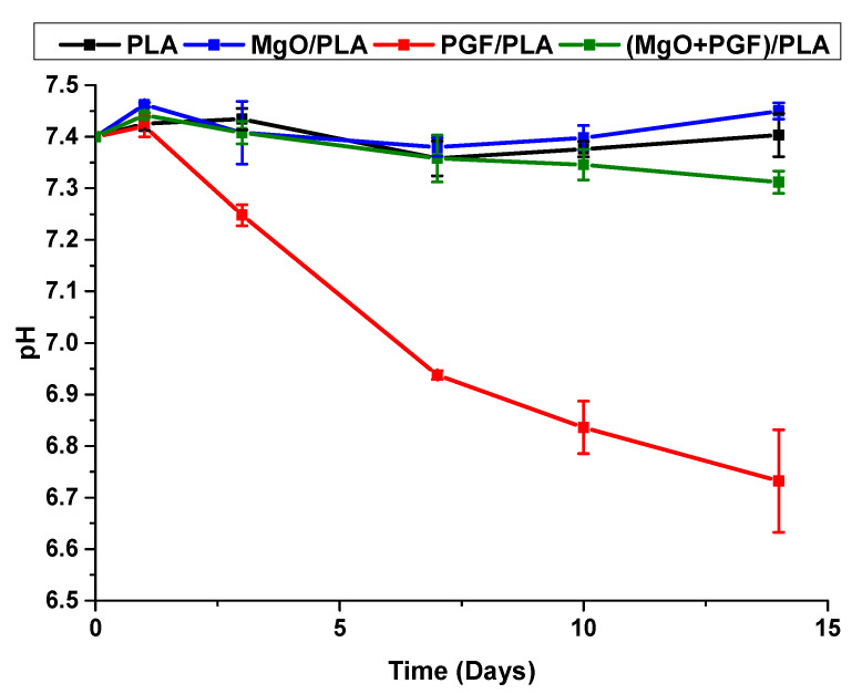
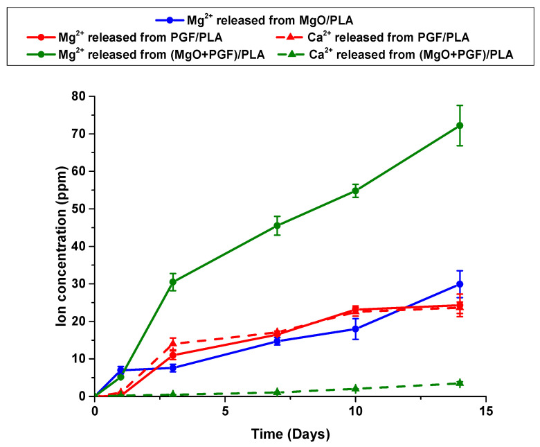
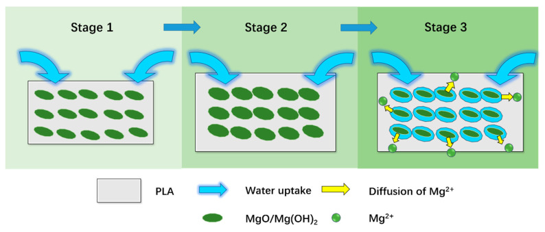
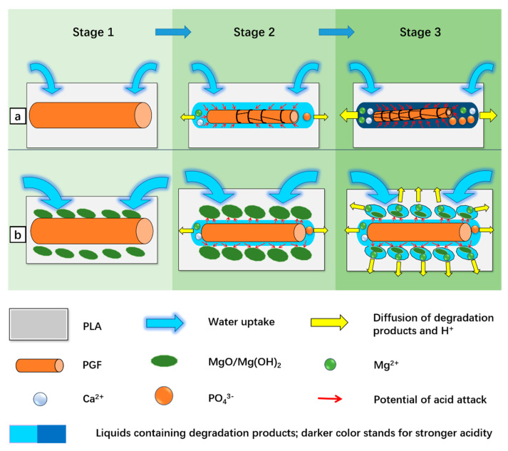
Composites of biodegradable phosphate glass fiber and polylactic acid (PGF/PLA) show potential for bone tissue engineering scaffolds, due to their ability to release Ca, P, and Mg during degradation, thus promoting the bone repair. Nevertheless, glass degradation tends to acidify the surrounding aqueous environment, which may adversely affect the viability and bone-forming activities of osteoblasts. In this work, MgO was investigated as a neutralizing agent. Porous network-phase gyroid scaffolds were additive-manufactured using four different materials: PLA, MgO/PLA, PGF/PLA, and (MgO + PGF)/PLA. The addition of PGF enhanced compressive properties of scaffolds, and the resultant scaffolds were comparably strong and stiff with human trabecular bone. While the degradation of PGF/PLA composite induced considerable acidity in degradation media and intensified the degradation of PGF in return, the degradation media of (MgO + PGF)/PLA maintained a neutral pH close to a physiological environment. The experiment results indicated the possible mechanism of MgO as the neutralizing agent: the local acidity was buffered as the MgO reacted with the acidic degradation products thereby inhibiting the degradation of PGF from being intensified in an acidic environment. The (MgO + PGF)/PLA composite scaffold appears to be a candidate for bone tissue engineering.

Keywords: additive manufacturing; bone tissue engineering scaffold; gyroid; phosphate glass fiber; polylactic acid.


Conflict of interest statement

The authors declare no conflict of interest.

Figures

Figure 1
Figure 1
(a) Gyroid scaffolds intended for the compressive test, with the 12 mm × 25 mm plane of scaffolds was placed flat on the build plate; (b) Gyroid scaffold intended for in vitro degradation. The models were displayed in the Simplify3D slicing software, with the width of square grids equals to 1 cm.
Figure 2
Figure 2
(a) Gyroid scaffolds intended for the compressive test; (b) Gyroid scaffolds intended for in-vitro degradation study. The material compositions of the four scaffolds (from left to right) are PLA, MgO/PLA, PGF/PLA, and (MgO + PGF)/PLA, respectively.
Figure 3
Figure 3
(a) Photograph of gyroid scaffolds before and after compression tests. Cracks propagated along the interlayer boundaries were highlighted with ovals; (b) Typical stress–strain curves of the gyroid scaffolds obtained from the compressive tests; (c) Summary of compressive strength and compressive modulus of the scaffold, with error bars representing the standard deviation. Five replicates were tested for each material. “p < 0.05” indicates statistical significance as analyzed based on unpaired t-tests.
Figure 4
Figure 4
Distribution of the length of PGFs in PGF/PLA and (MgO + PGF)/PLA composite scaf.
Figure 5
Figure 5
Photograph of scaffolds after 14 days of degradation.
Figure 6
Figure 6
The freeze-fractured surfaces of different scaffolds before and after 14 days of in vitro degradation in Tris-HCl under 37 °C.
Figure 7
Figure 7
SEM image of the top surface of different scaffolds before and after 14 days of in vitro degradation in Tris-HCl under 37 °C.
Figure 8
Figure 8
Photograph showing the yellow sediments in the degradation media immersed with PGF/PLA for 14 days, and the SEM images as well as the Energy-dispersive X-ray Spectroscopy (EDS) spectra of both the sediments and the top surfaces of PGF/PLA composite scaffolds.
Figure 9
Figure 9
Water uptake profile of scaffolds during 14 days of in vitro degradation in Tris-HCl buffer under 37 °C.
Figure 10
Figure 10
Mass change profile of scaffolds during 14 days of in vitro degradation in Tris-HCl buffer under 37 °C.
Figure 11
Figure 11
pH profile of the Tris-HCl immersed with different scaffolds during 14 days of in vitro degradation.
Figure 12
Figure 12
Ion concentration profile in the degradation media immersed with different scaffolds.
Figure 13
Figure 13
Schematic representation of the degradation of MgO/PLA composite scaffolds.
Figure 14
Figure 14
Schematic representation of the possible degradation mechanisms for different composites. (a) a mechanism for the binary PGF/PLA composite; (b) mechanism for the ternary (MgO + PGF/PLA) composite.

Similar articles

Cited by

References

    1. Williams D., Zhang X., editors. Definitions of Biomaterials for the Twenty-First Century. Elsevier; Amsterdam, The Netherlands: 2019. VI—Regenerative medicine; pp. 115–153. - DOI
    1. Amini A.R., Laurencin C.T., Nukavarapu S.P. Bone Tissue Engineering: Recent Advances and Challenges. Crit. Rev. Biomed. Eng. 2012;40:363–408. doi: 10.1615/CritRevBiomedEng.v40.i5.10. - DOI - PMC - PubMed
    1. Bose S., Vahabzadeh S., Bandyopadhyay A. Bone tissue engineering using 3D printing. Mater. Today. 2013;16:496–504. doi: 10.1016/j.mattod.2013.11.017. - DOI
    1. Hollister S.J. Porous scaffold design for tissue engineering. Nat. Mater. 2005;4:518–524. doi: 10.1038/nmat1421. - DOI - PubMed
    1. Vijayavenkataraman S., Zhang L., Zhang S., Hsi Fuh J.Y., Lu W.F. Triply Periodic Minimal Surfaces Sheet Scaffolds for Tissue Engineering Applications: An Optimization Approach toward Biomimetic Scaffold Design. ACS Appl. Bio Mater. 2018;1:259–269. doi: 10.1021/acsabm.8b00052. - DOI - PubMed

LinkOut - more resources